©The Author(s) 2017.
World J Gastroenterol. Mar 7, 2017; 23(9): 1725-1734
Published online Mar 7, 2017. doi: 10.3748/wjg.v23.i9.1725
Published online Mar 7, 2017. doi: 10.3748/wjg.v23.i9.1725
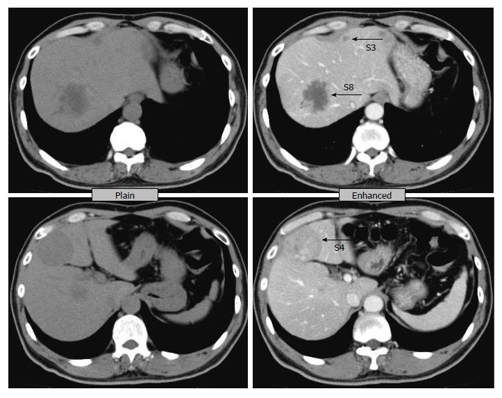
Figure 1 Computed tomography before treatment.
An abdominal computed tomography scan revealed liver tumors in Segment 3, Segment 4, and Segment 8. The tumor in Segment 8 is hypodense with peripheral enhancement. The tumor in Segment 4 is a well-defined isodense tumor with homogeneous enhancement.
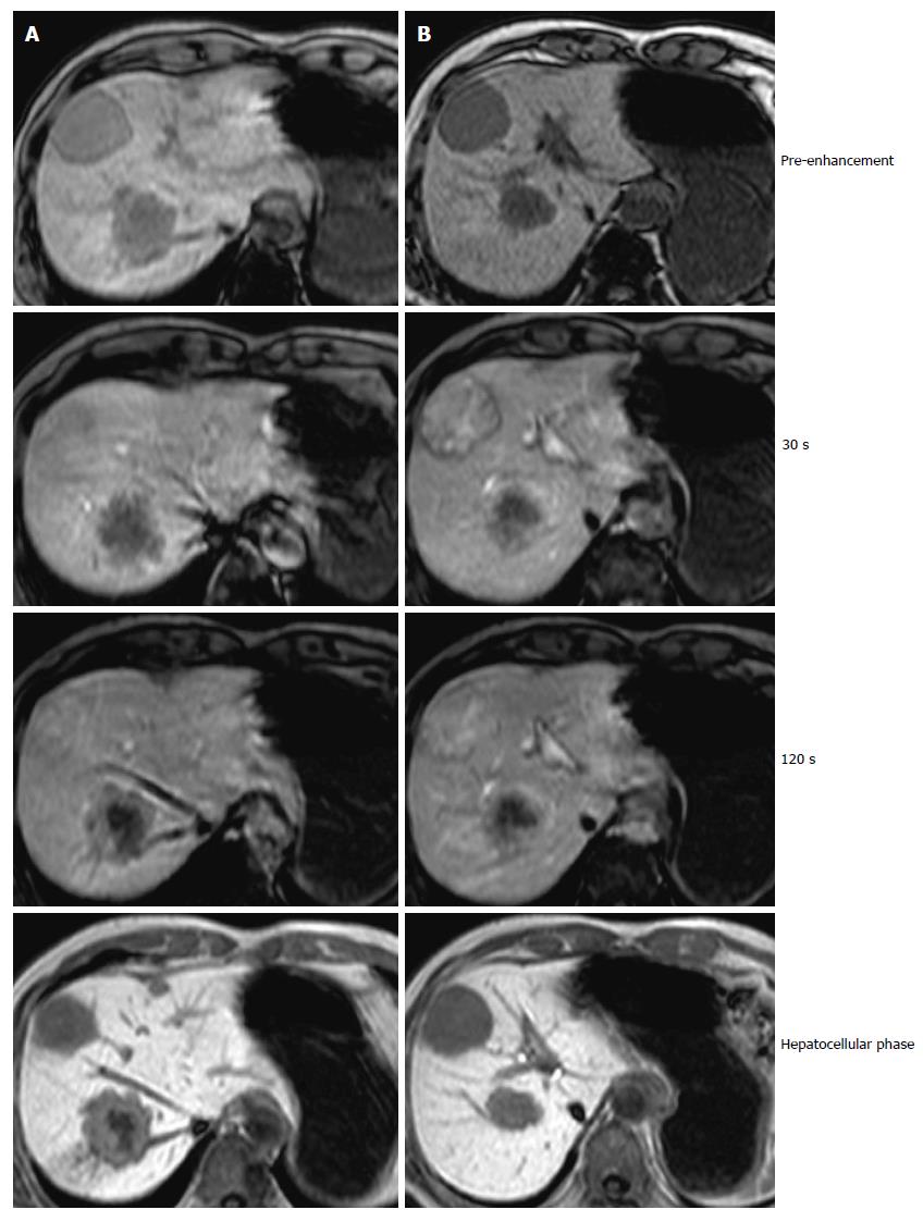
Figure 2 Ethoxibenzyl-magnetic resonance imaging before treatment.
The tumors in Segments 3 and 8 showed gradual peripheral enhancement, while the tumor in Segment 4 showed heterogeneous enhancement and washout characteristics. A: S3 and S8 gradual peripheral enhancement; B: S4 heterogeneous enhancement.
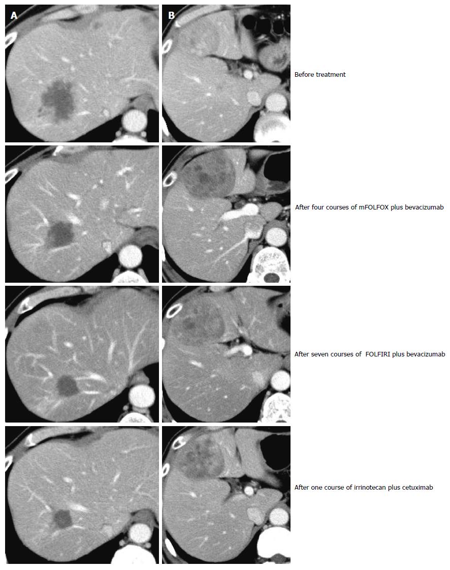
Figure 3 Changes in computed tomography images during treatment.
The tumors in Segments 3 and 8 showed a gradual decrease in size, while the tumor in Segment 4 exhibited a one-time increase and then a decrease in size. A: S8 gradually decreased in size/S3 disappeared; B: S4 once increased then decreased in size.
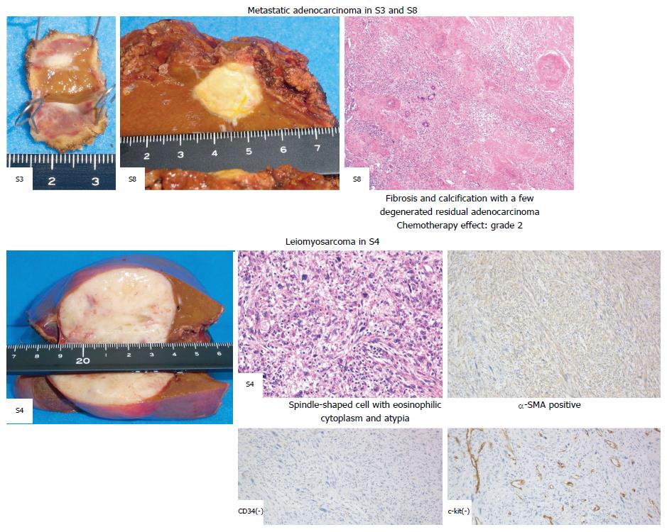
Figure 4 Pathological diagnoses of live lesions.
The tumors in Segments 3 and 8 revealed fibrosis and calcification, with a few degenerated residual adenocarcinomas, while the tumor in Segment 4 consisted of irregular fascicles of spindle-shaped cells and was positive for SMA and negative for CD34 and c-kit. SMA: Smooth muscle actin.
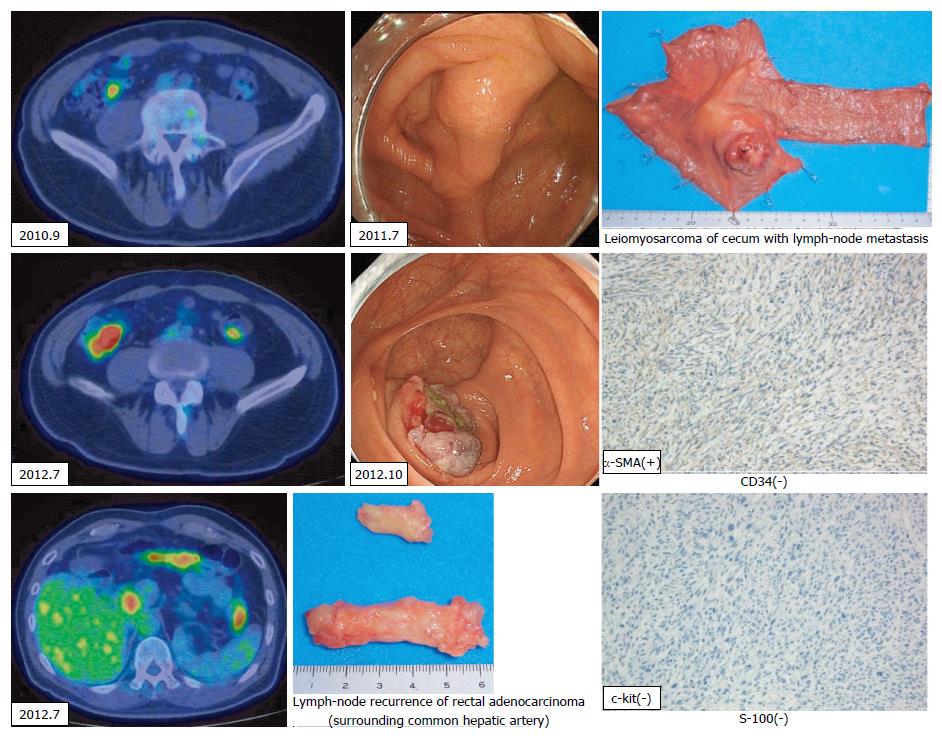
Figure 5 Cecal leiomyosarcoma and lymph-node recurrence.
A cecal tumor and lymph-node swelling around the common hepatic artery were discovered in a positron emission tomography-computed tomography (PET-CT) scan. Retrospectively, accumulation had been observed in the cecum in a PET-CT scan in September 2010, and a submucosal tumor was suspected based on a colonoscopy taken in July 2011. SMA: Smooth muscle actin.
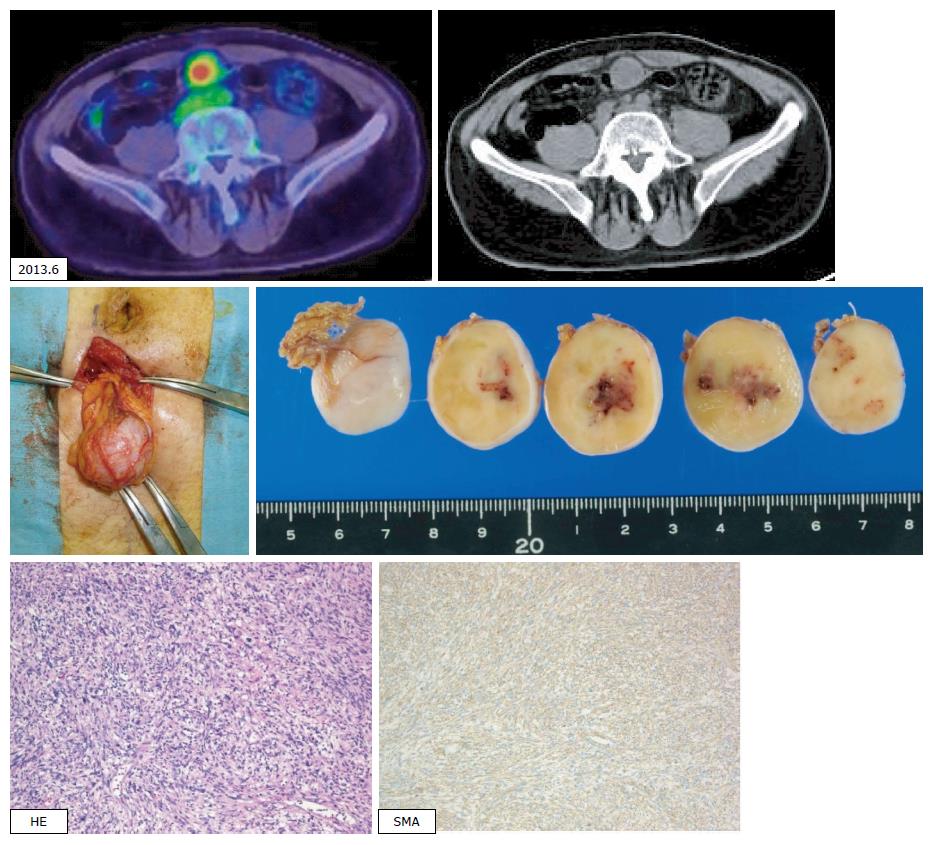
Figure 6 Peritoneal recurrences.
A tumor just below the peritoneum was discovered. As the accumulation was recognized in a positron emission tomography-computed tomography scan and no other accumulation was observed, extirpation of the tumor was carried out. Pathological diagnosis was leiomyosarcoma of the omentum, compatible with recurrence. SMA: Smooth muscle actin.
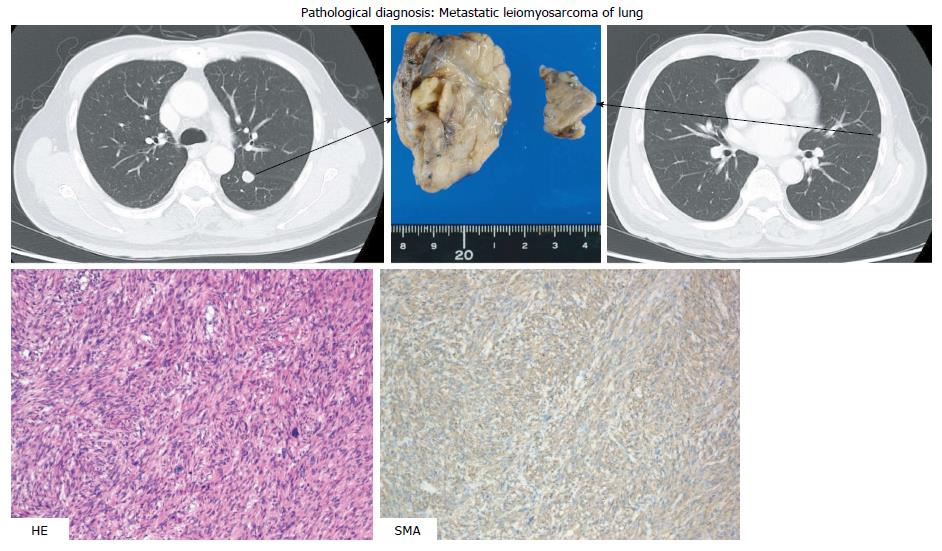
Figure 7 Lung recurrences.
In a chest CT scan, two coin lesions were discovered in the left lung. As lung metastases were strongly suspected, partial resections of the left upper lobe and left lower lobe were performed. Pathological diagnosis was metastatic leiomyosarcoma of the lung.
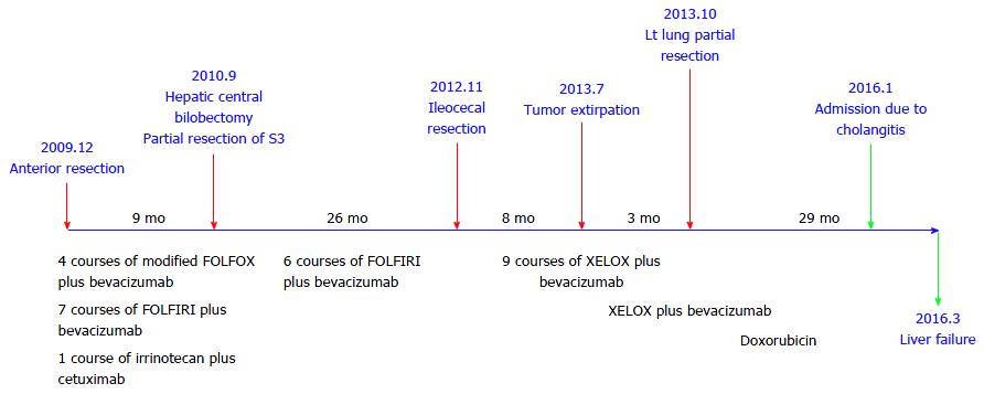
Figure 8 Clinical courses.
- Citation: Aoki H, Arata T, Utsumi M, Mushiake Y, Kunitomo T, Yasuhara I, Taniguchi F, Katsuda K, Tanakaya K, Takeuchi H, Yamasaki R. Synchronous coexistence of liver metastases from cecal leiomyosarcoma and rectal adenocarcinoma: A case report. World J Gastroenterol 2017; 23(9): 1725-1734
- URL: https://www.wjgnet.com/1007-9327/full/v23/i9/1725.htm
- DOI: https://dx.doi.org/10.3748/wjg.v23.i9.1725


