©The Author(s) 2017.
World J Gastroenterol. Mar 7, 2017; 23(9): 1720-1724
Published online Mar 7, 2017. doi: 10.3748/wjg.v23.i9.1720
Published online Mar 7, 2017. doi: 10.3748/wjg.v23.i9.1720
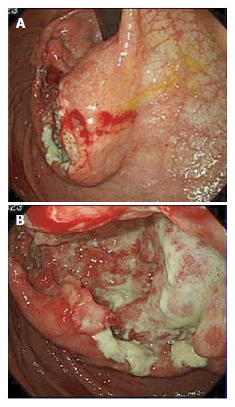
Figure 1 Gastroscopy revealed a large tumor with ulceration in the upper body of the stomach.
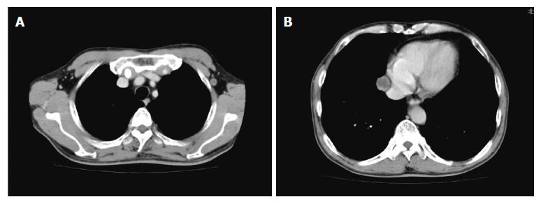
Figure 2 Computed tomography reveals a tumor in the left axilla (A, diameter: 1 cm) and a tumor in the right mediastinum (B, diameter: 2 cm).
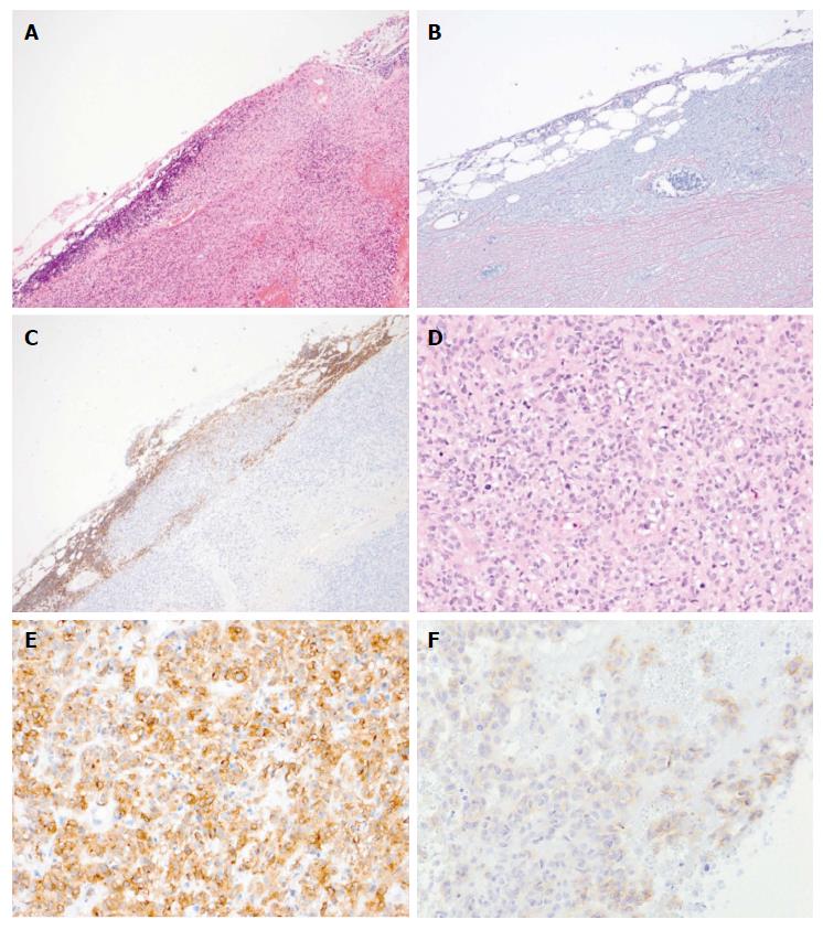
Figure 3 Pathological findings of the biopsied left axilla lymph node.
Analysis of the tumor revealed tunicate formation and the survival of lymphoid tissue [hematoxylin and eosin staining(A), silver impregnation (B), and Leukocyte common antigen (C) (magnification × 40)]. The tumor exhibited monotonous spindle cells (D, hematoxylin and eosin staining), and the cells were positive for c-kit (E) and DOG1 (F, magnification × 100).
- Citation: Kubo N, Takeuchi N. Gastrointestinal stromal tumor of the stomach with axillary lymph node metastasis: A case report. World J Gastroenterol 2017; 23(9): 1720-1724
- URL: https://www.wjgnet.com/1007-9327/full/v23/i9/1720.htm
- DOI: https://dx.doi.org/10.3748/wjg.v23.i9.1720


