©The Author(s) 2017.
World J Gastroenterol. Mar 7, 2017; 23(9): 1712-1719
Published online Mar 7, 2017. doi: 10.3748/wjg.v23.i9.1712
Published online Mar 7, 2017. doi: 10.3748/wjg.v23.i9.1712
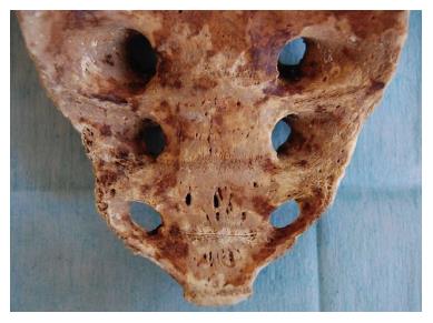
Figure 1 Sacrum specimen.
Multiple sacral basivertebral vein foramin, between 2-4 mm, are seen on S4-S5.
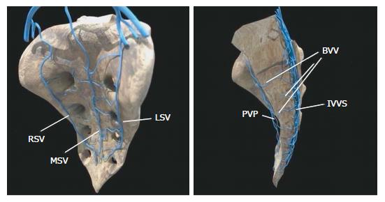
Figure 2 Diagram showing the sacral venous system.
RSV: Right sacral vein; LSV: Left sacral vein; MSV: Middle sacral vein; PVP: Presacral venous plexus; IVVS: Internal vertebral venous system; BVV: Basivertebral vein.
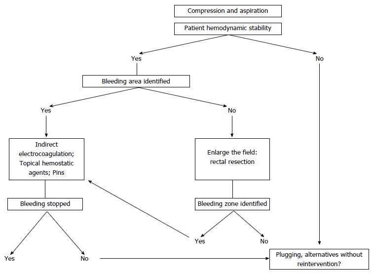
Figure 3 Presacral venous hemorrhaging: treatment algorithm.
- Citation: Casal Núñez JE, Vigorita V, Ruano Poblador A, Gay Fernández AM, Toscano Novella M&, Cáceres Alvarado N, Pérez Dominguez L. Presacral venous bleeding during mobilization in rectal cancer. World J Gastroenterol 2017; 23(9): 1712-1719
- URL: https://www.wjgnet.com/1007-9327/full/v23/i9/1712.htm
- DOI: https://dx.doi.org/10.3748/wjg.v23.i9.1712


