©The Author(s) 2017.
World J Gastroenterol. Dec 28, 2017; 23(48): 8544-8552
Published online Dec 28, 2017. doi: 10.3748/wjg.v23.i48.8544
Published online Dec 28, 2017. doi: 10.3748/wjg.v23.i48.8544
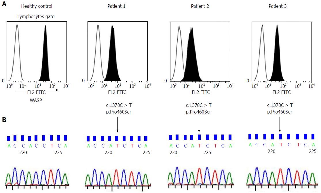
Figure 1 Analysis of Wiskott-Aldrich syndrome protein in patients’ lymphocytes (A).
Histograms represent intracellular Wiskott-Aldrich syndrome protein (WASP) staining (shaded area) and negative staining (unshaded area). Three patients with childhood-onset IBD presented low expression of WASP compared with that in the healthy control. B: WAS gene analysis. The three patients who showed low expression of WASP had the same mutation, c.1378C>T p.Pro460Ser.
Figure 2 Cutaneous manifestations of Patient 1 (scaling eczema and pigmentation).
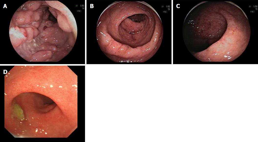
Figure 3 Endoscopic findings in the patients with WAS c.
1378C>T, p.Pro460Ser mutation. A: Patient 1: long linear ulcerations and cobble stone appearance in the ileum. B and C: Patient 2: edematous and friable mucosa with superficial bleeding in the descending colon and sigmoid colon. D: Patient 3: edematous mucosa with granularity and erythema in the rectum.
- Citation: Ohya T, Yanagimachi M, Iwasawa K, Umetsu S, Sogo T, Inui A, Fujisawa T, Ito S. Childhood-onset inflammatory bowel diseases associated with mutation of Wiskott-Aldrich syndrome protein gene. World J Gastroenterol 2017; 23(48): 8544-8552
- URL: https://www.wjgnet.com/1007-9327/full/v23/i48/8544.htm
- DOI: https://dx.doi.org/10.3748/wjg.v23.i48.8544


