©The Author(s) 2017.
World J Gastroenterol. Nov 28, 2017; 23(44): 7875-7880
Published online Nov 28, 2017. doi: 10.3748/wjg.v23.i44.7875
Published online Nov 28, 2017. doi: 10.3748/wjg.v23.i44.7875
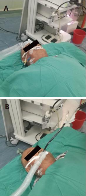
Figure 1 The patients were placed in the supine position.
Electrocardiography, noninvasive arterial blood pressure monitoring, and pulse oximetry were performed as a part of the standard monitoring protocol. Sufentanil (0.5-1 μg/kg) was injected to induce mild sedation and restrain the stress response to intubation. Anesthesia was induced with 1-2 mg/kg of propofol and maintained with 2-5 mg/kg per hour of propofol. Scoline and cisatracurium were injected for the endotracheal intubation.
Figure 2 Endoscopic injection sclerotherapy for esophageal varices.
A: The patient was EV grade ≥ F2 and had a severe red color sign, according to endoscopic findings; B: Endoscopic injection sclerotherapy (EIS) was performed with a standard endoscope (SV-290; Olympus Corporation; Tokyo; Japan). The endoscopic puncture needle used in EIS was a 23-gauge Varixer needle; C: The endoscopic finding after EIS.
- Citation: Yu Y, Qi SL, Zhang Y. Role of combined propofol and sufentanil anesthesia in endoscopic injection sclerotherapy for esophageal varices. World J Gastroenterol 2017; 23(44): 7875-7880
- URL: https://www.wjgnet.com/1007-9327/full/v23/i44/7875.htm
- DOI: https://dx.doi.org/10.3748/wjg.v23.i44.7875


