Copyright
©The Author(s) 2017.
World J Gastroenterol. Jan 28, 2017; 23(4): 735-739
Published online Jan 28, 2017. doi: 10.3748/wjg.v23.i4.735
Published online Jan 28, 2017. doi: 10.3748/wjg.v23.i4.735
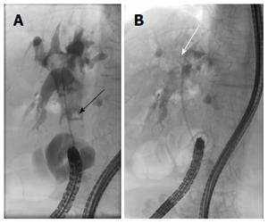
Figure 1 Endoscopic retrograde cholangiopancreatography image.
A: Stones in common bile duct (black arrow); B: The plastic stent had been placed improperly, and the edge was coiled around in the common bile duct (white arrow). There was no sign of collapse or leakage in the biliary tract.
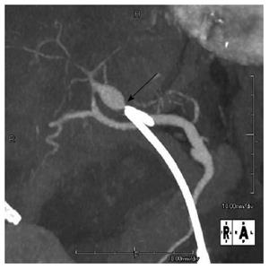
Figure 2 Abdominal computed tomography angiography.
A 13 mm × 10 mm pseudoaneurysm was found at the proximal anterior segment of the right hepatic artery; The hepatic side edge of the plastic stent (black arrow) was close to the pseudoaneurysm.
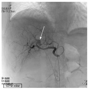
Figure 3 The pseudoaneurysm was detected upon celiac axis injection, with the aneurysmal sac slightly lateral to the edge of the plastic stent (arrow).
The sac was no longer seen on a selective digital subtraction angiography image after successful embolization. Blood flow was maintained in the anterior segment of the right hepatic artery.
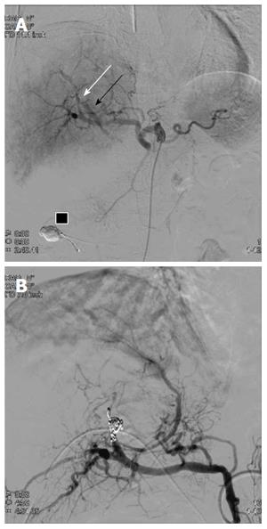
Figure 4 The 2nd digital subtraction angiography image.
A: Selective digital subtraction angiography image of the common hepatic artery showing an arterio-biliary fistula (white arrow) between A8 and the common bile duct (black arrow). Embolic material that migrated into the afferent loop is present (black box); B: Proximal A8 embolization obliterated the pseudoaneurysm. Leakage of contrast medium was completely controlled after embolization.
- Citation: Yasuda M, Sato H, Koyama Y, Sakakida T, Kawakami T, Nishimura T, Fujii H, Nakatsugawa Y, Yamada S, Tomatsuri N, Okuyama Y, Kimura H, Ito T, Morishita H, Yoshida N. Late-onset severe biliary bleeding after endoscopic pigtail plastic stent insertion. World J Gastroenterol 2017; 23(4): 735-739
- URL: https://www.wjgnet.com/1007-9327/full/v23/i4/735.htm
- DOI: https://dx.doi.org/10.3748/wjg.v23.i4.735


