Copyright
©The Author(s) 2017.
World J Gastroenterol. Jan 28, 2017; 23(4): 661-667
Published online Jan 28, 2017. doi: 10.3748/wjg.v23.i4.661
Published online Jan 28, 2017. doi: 10.3748/wjg.v23.i4.661
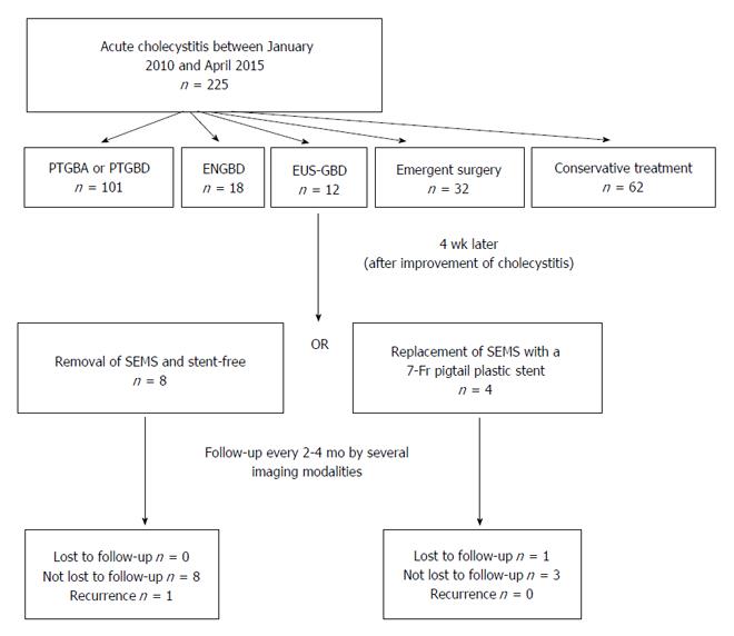
Figure 1 Strategy of endoscopic ultrasound-guided gallbladder drainage procedure.
ENGBD: Endoscopic naso-gallbladder drainage; EUS-GBD: Endoscopic ultrasound-guided gallbladder drainage; PTGBA: Percutaneous transhepatic gallbladder aspiration; PTGBD: Percutaneous transhepatic gallbladder drainage; SEMS: Self-expandable metal stent.
Figure 2 Endosonographic image.
A: Endosonographic image before gallbladder drainage. Contents in the gallbladder were mainly sludge without apparent gallstones; B: Endosonographic image at the time of recurrence of cholecystitis. Sludge volume in the gallbladder was more increased than when gallbladder drainage was performed.
Figure 3 Esophagogastroduodenoscopy and computed tomography.
A: Esophagogastroduodenoscopy image of deployment of the metal stent in the duodenum; B: Esophagogastroduodenoscopy image of the fistula 1 wk after removal of the metal stent; C: Computed tomography after removal of the metal stent showing air image in the gallbladder.
- Citation: Kamata K, Takenaka M, Kitano M, Omoto S, Miyata T, Minaga K, Yamao K, Imai H, Sakurai T, Watanabe T, Nishida N, Kudo M. Endoscopic ultrasound-guided gallbladder drainage for acute cholecystitis: Long-term outcomes after removal of a self-expandable metal stent. World J Gastroenterol 2017; 23(4): 661-667
- URL: https://www.wjgnet.com/1007-9327/full/v23/i4/661.htm
- DOI: https://dx.doi.org/10.3748/wjg.v23.i4.661


