©The Author(s) 2017.
World J Gastroenterol. Oct 21, 2017; 23(39): 7119-7128
Published online Oct 21, 2017. doi: 10.3748/wjg.v23.i39.7119
Published online Oct 21, 2017. doi: 10.3748/wjg.v23.i39.7119
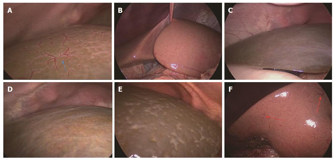
Figure 1 Laparoscopic images of the liver surface in the retrospective study.
A: HSST sign in a 70-day-old boy with biliary atresia (blue arrow); B: Image of a 64-day-old boy with Hirschsprung’s disease; C: Image of an 82-day-old boy with infantile hepatitis; D: Image of a 72-day-old girl with biliary hypoplasia; E: Image of a 70-day-old boy with total parenteral nutrition-induced cholestatic cirrhosis; F: Small vessel plexuses (red arrows) observed in a 55-day-old boy with cytomegalovirus hepatitis. The HSST sign does not exist in the images B-E. HSST: Hepatic subcapsular spider-like telangiectasis.
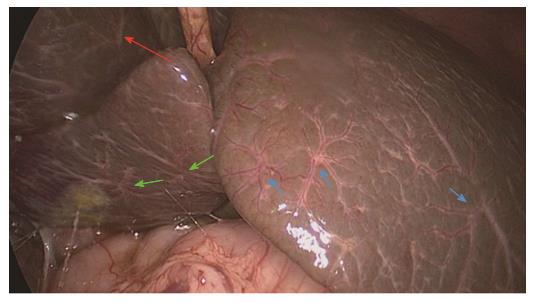
Figure 2 Hepatic subcapsular spider-like telangiectasis sign in a 68-day-old boy with biliary atresia.
A hepatic subcapsular spider-like telangiectasis sign was widely distributed on the liver surface, including on the visceral facies of the right liver lobe (red arrow), on diaphragmatic facies of the left liver lobe (blue arrows) and on the surface of the caudate lobe (green arrows).
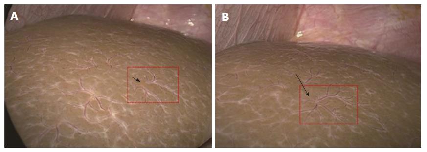
Figure 3 Different types of hepatic subcapsular spider-like telangiectasis signs.
A: Dispersed type (short arrow) of the HSST sign in a 75-day-old boy with BA; B: Concentrated type (long arrow) of the HSST sign in a 77-day-old boy with BA. HSST: Hepatic subcapsular spider-like telangiectasis; BA: Biliary atresia.
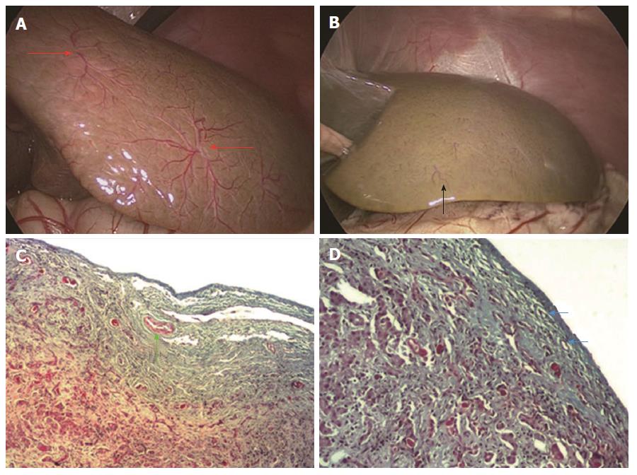
Figure 4 Hepatic subcapsular spider-like telangiectasis sign and pathologic evaluation in the validation set.
A: Concentrated type of the HSST sign (red arrow) in a 68-day-old boy with BA; B: Vessels (black arrow) in biliary hypoplasia; C: Vessels in (A) was revealed as dilated small arteries (green arrow) in the hepatic subcapsular area; D: Vessels in biliary hypoplasia (B) was revealed as dilated capillaries (blue arrow) in the hepatic subcapsular area. (Trichrome staining; ×100). HSST: Hepatic subcapsular spider-like telangiectasis; BA: Biliary atresia.
Figure 5 Laparoscopic images of hepatic subcapsular spider-like telangiectasis sign in biliary atresia patients at different ages.
A: HSST sign (red arrow) in a 40-day-old boy with BA. There was no cirrhosis at gross inspection; B: HSST sign (blue arrow) in a 156-day-old boy with BA, who had hepatic cirrhosis with ascites and underwent liver transplantation. The HSST sign is obvious in both patients. HSST: Hepatic subcapsular spider-like telangiectasis; BA: Biliary atresia.
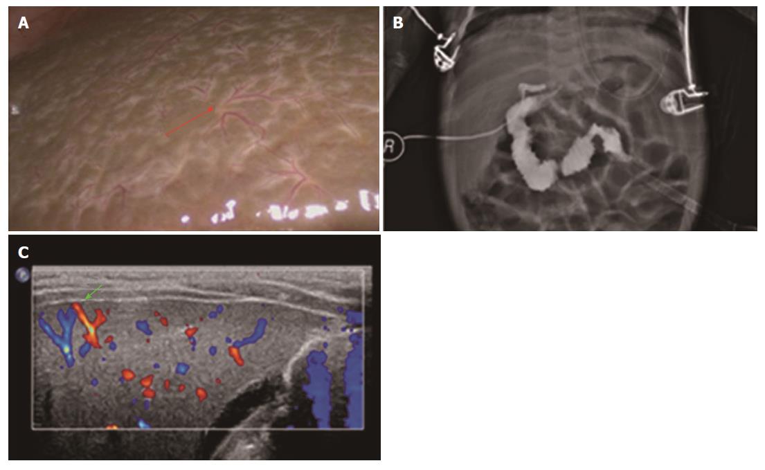
Figure 6 Images in a 53-day-old patient with biliary atresia.
A: Dispersed type of the HSST sign (red arrow); B: Image of laparoscopic cholangiography indicates biliary atresia; C: Hepatic arterial flow extending to the hepatic surface on color Doppler US image (green arrow) indicates positive HSF. Laparoscopic HSST sign and the result of laparoscopic cholangiography were easier to identify than distinguishing HSF in a color Doppler US image. HSST: Hepatic subcapsular spider-like telangiectasis; US: Ultrasonography; HSF: Hepatic subcapsular flow.
- Citation: Zhou Y, Jiang M, Tang ST, Yang L, Zhang X, Yang DH, Xiong M, Li S, Cao GQ, Wang Y. Laparoscopic finding of a hepatic subcapsular spider-like telangiectasis sign in biliary atresia. World J Gastroenterol 2017; 23(39): 7119-7128
- URL: https://www.wjgnet.com/1007-9327/full/v23/i39/7119.htm
- DOI: https://dx.doi.org/10.3748/wjg.v23.i39.7119


