©The Author(s) 2017.
World J Gastroenterol. Sep 7, 2017; 23(33): 6187-6193
Published online Sep 7, 2017. doi: 10.3748/wjg.v23.i33.6187
Published online Sep 7, 2017. doi: 10.3748/wjg.v23.i33.6187
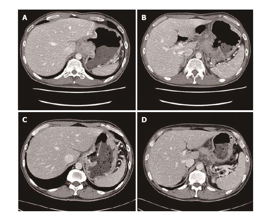
Figure 1 On admission, computed tomography revealed a bulky ulcerative mass with direct invasion of pancreas and left diaphragm (A and B).
After 3 cycle of neoadjuvant chemotherapy (capecitabine and oxaliplatin), the mass had markedly decrease (C and D).
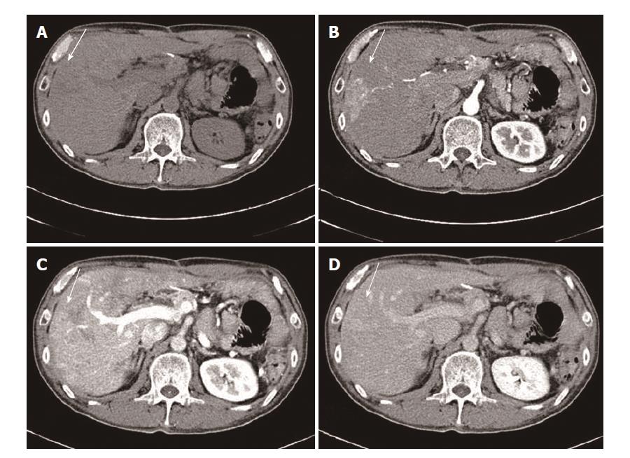
Figure 2 After total six cycle of chemotherapy (capecitabine and oxaliplatin), dynamic computed tomography showed an arterial enhanced mass like lesion.
A: Pre-enhanced image; B: Arterial phase image; C: Portal phase image; D: Delayed phase image.
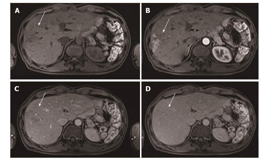
Figure 3 Dynamic magnetic resonance imaging demonstrated an arterial enhance mass like lesion as same as computed tomography scan.
A: Pre-enhanced image; B: Arterial phase image; C: Portal phase image; D: delayed phase image.
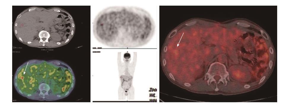
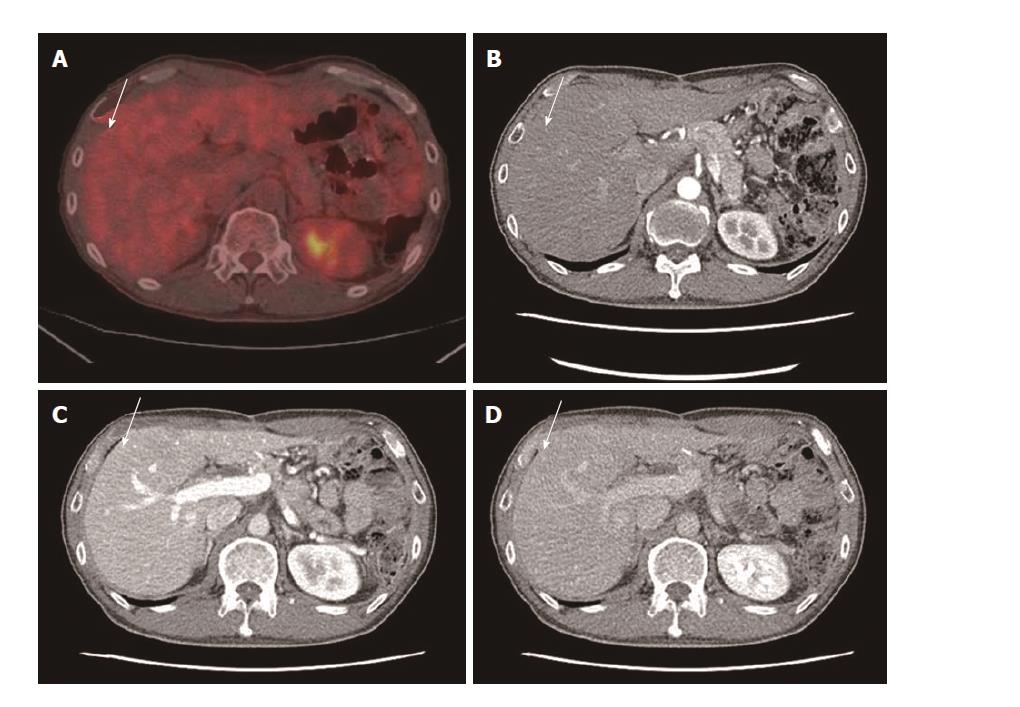
Figure 4 This arterial enhances mass like lesion had a hypermetabolism in positron emission tomography computed tomography.
Figure 5 After adjuvant chemotherapy without oxaliplatin, the arterial enhance lesion disappeared.
A: PET CT; B: Arterial phase image; C: Portal phase image; D: Delayed phase image.
- Citation: Kim HB, Park SG. Arterioportal shunt incidental to treatment with oxaliplatin that mimics recurrent gastric cancer. World J Gastroenterol 2017; 23(33): 6187-6193
- URL: https://www.wjgnet.com/1007-9327/full/v23/i33/6187.htm
- DOI: https://dx.doi.org/10.3748/wjg.v23.i33.6187


