Copyright
©The Author(s) 2017.
World J Gastroenterol. Aug 28, 2017; 23(32): 5962-5968
Published online Aug 28, 2017. doi: 10.3748/wjg.v23.i32.5962
Published online Aug 28, 2017. doi: 10.3748/wjg.v23.i32.5962
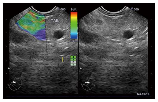
Figure 1 A patient with chronic pancreatitis showing heterogeneous soft tissue (green, yellow, and red), and interpreted as fibrosis or inflammation.
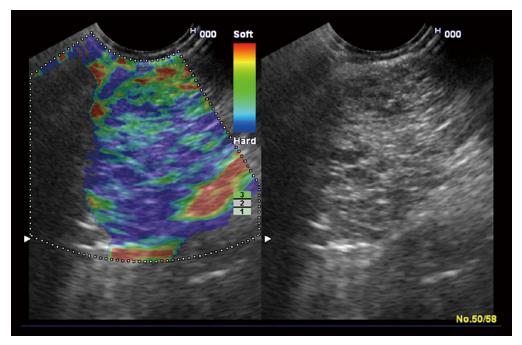
Figure 2 A patient with elasticity score 3 showing mixed hard and soft tissues (mixed colors) or a honeycombed elastography pattern, interpreted as indeterminate for malignancy.
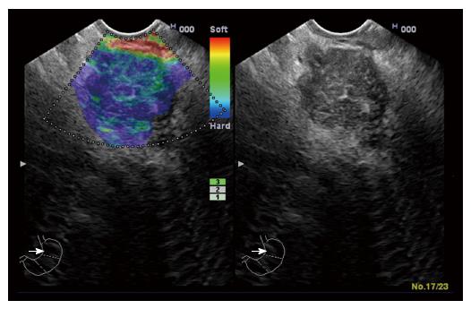
Figure 3 A patient with autoimmune pancreatitis showing elasticity score 3.
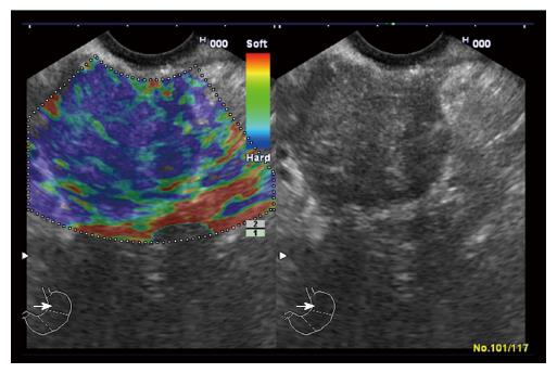
Figure 4 A patient with advanced malignant lesions with necrotic areas (elasticity score 5) showing predominantly hard (blue) lesion with dispersed heterogenic soft (green) areas.
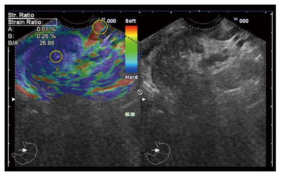
Figure 5 A patient with pancreatic head malignancy showing high stain ratio (25.
86).
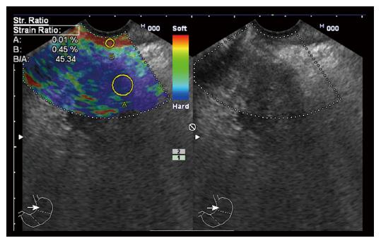
Figure 6 A patient with pancreatic head malignancy showing very high stain ratio (45.
34).
- Citation: Okasha H, Elkholy S, El-Sayed R, Wifi MN, El-Nady M, El-Nabawi W, El-Dayem WA, Radwan MI, Farag A, El-sherif Y, Al-Gemeie E, Salman A, El-Sherbiny M, El-Mazny A, Mahdy RE. Real time endoscopic ultrasound elastography and strain ratio in the diagnosis of solid pancreatic lesions. World J Gastroenterol 2017; 23(32): 5962-5968
- URL: https://www.wjgnet.com/1007-9327/full/v23/i32/5962.htm
- DOI: https://dx.doi.org/10.3748/wjg.v23.i32.5962


