Copyright
©The Author(s) 2017.
World J Gastroenterol. Aug 28, 2017; 23(32): 5836-5848
Published online Aug 28, 2017. doi: 10.3748/wjg.v23.i32.5836
Published online Aug 28, 2017. doi: 10.3748/wjg.v23.i32.5836
Figure 1 Diffuse episcleritis.
A: Superior view; B: Episcleral injection at slit lamp exam; C: Inferior view. Personal archive.
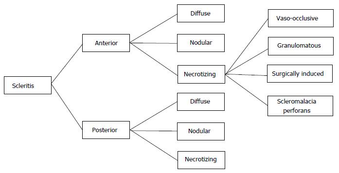
Figure 2 Classification of scleritis[64].
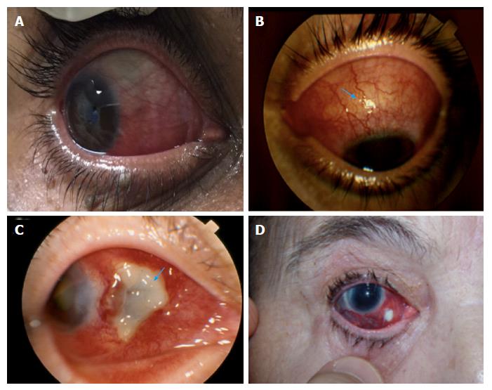
Figure 3 Clinical presentation of scleritis.
A: Anterior diffuse scleritis (personal archive); B: Anterior nodular scleritis (personal archive). The differential diagnosis is based on the presence of a sclera nodule (arrow); C: Anterior necrotizing scleritis, showing the avascular area of necrosis (arrow) (personal archive); D: Anterior necrotizing surgically-induced scleritis, induced by scleral biopsy (courtesy of Prof. Andre Curi).
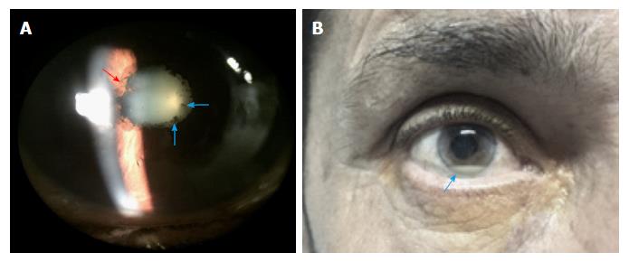
Figure 4 Anterior uveitis.
A: Slit lamp exam revealed posterior synechiae (red arrow) and pigment deposits on the anterior lens capsule (blue arrow) (personal archive); B: Inflammatory cells in the anterior chamber of the eye causing hypopyon (arrow) (personal archive).
- Citation: Troncoso LL, Biancardi AL, de Moraes Jr HV, Zaltman C. Ophthalmic manifestations in patients with inflammatory bowel disease: A review. World J Gastroenterol 2017; 23(32): 5836-5848
- URL: https://www.wjgnet.com/1007-9327/full/v23/i32/5836.htm
- DOI: https://dx.doi.org/10.3748/wjg.v23.i32.5836


