Copyright
©The Author(s) 2017.
World J Gastroenterol. Jul 7, 2017; 23(25): 4579-4586
Published online Jul 7, 2017. doi: 10.3748/wjg.v23.i25.4579
Published online Jul 7, 2017. doi: 10.3748/wjg.v23.i25.4579
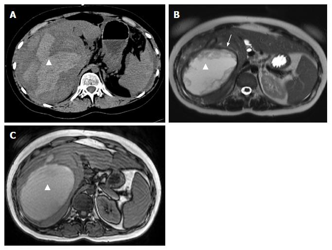
Figure 1 Representative case of grade II bleeding hepatocellular adenoma involving a 23-year-old female who presented at the emergency department with an acute hematoma in the right liver lobe.
A: Computed tomography without intravenous contrast shows a hyperdense fluid collection in the liver (triangle); B: Some months later after conservative treatment, the collection appeared hyperintense on T2-weighting, with a hemosiderin ring surrounding the collection (arrow); C: At the same time, the collection appeared hyperintense on T1-weighting.
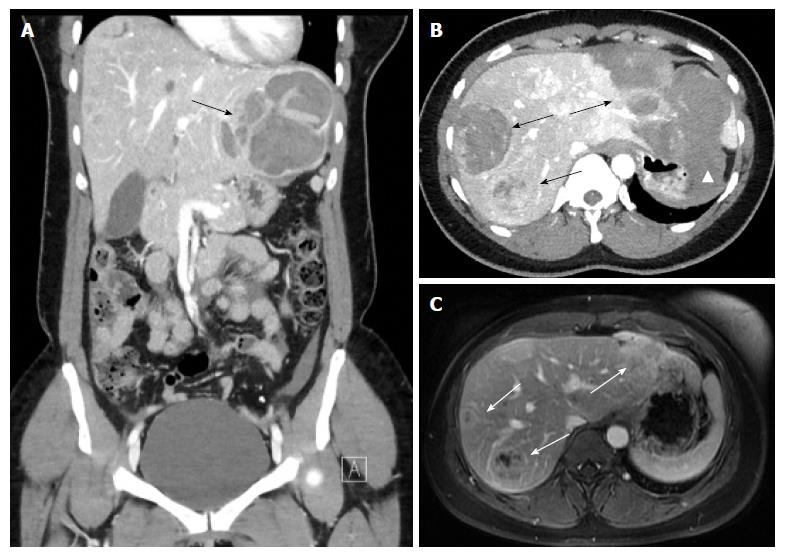
Figure 2 Representative case of grade III bleeding hepatocellular adenoma involving a 28-year-old female who presented at the emergency department.
A: Coronal computed tomography (CT) image in arterial phase shows a large hematoma in the left liver lobe (arrow); B: On the axial CT slice different bleeding adenomas are present in both lobes (arrows). Extracapsular peritoneal bleeding around the stomach is seen (triangle); C: One year later on magnetic resonance imaging in the venous phase all adenomas have decreased significantly in size.
- Citation: Klompenhouwer AJ, de Man RA, Thomeer MG, Ijzermans JN. Management and outcome of hepatocellular adenoma with massive bleeding at presentation. World J Gastroenterol 2017; 23(25): 4579-4586
- URL: https://www.wjgnet.com/1007-9327/full/v23/i25/4579.htm
- DOI: https://dx.doi.org/10.3748/wjg.v23.i25.4579


