©The Author(s) 2017.
World J Gastroenterol. Jun 21, 2017; 23(23): 4170-4180
Published online Jun 21, 2017. doi: 10.3748/wjg.v23.i23.4170
Published online Jun 21, 2017. doi: 10.3748/wjg.v23.i23.4170
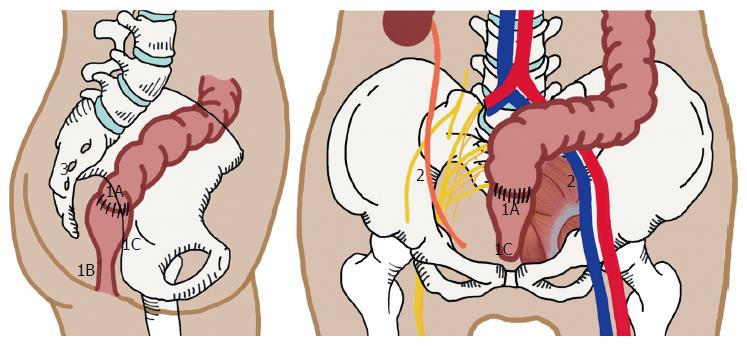
Figure 1 Patterns of pelvic recurrence.
1: Central; 1A: Anastomotic site; 1B: Perineal region, seen after abdominal perineal resection; 1C: Invasion to adjacent soft tissue involving genitourinary organs, or to pubic bone; 2: Lateral Pelvic Side Wall; 3: Posterior/Sacral Recurrence.
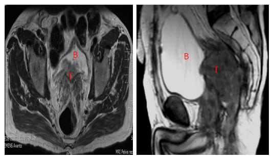
Figure 2 Central recurrence rectal cancer (T) involving the bladder (B).
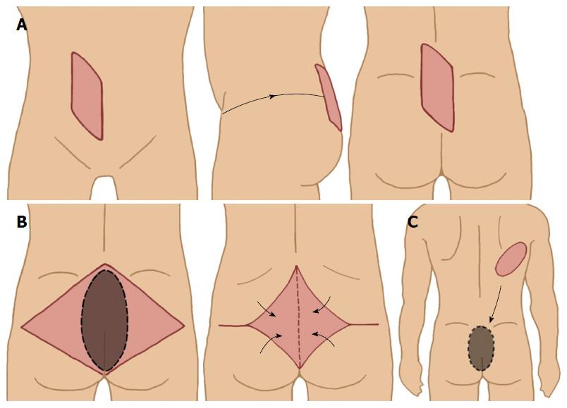
Figure 3 Various myocutaneous flaps used to cover perineal/sacral defect.
A: Vertical rectus abdominis myocutaneous flap; B: Gluteal muscle flap; C: Latissimus dorsi free flap.
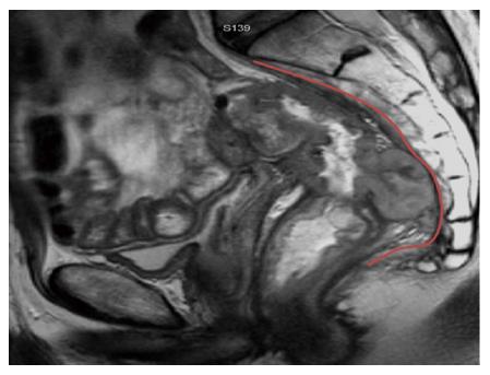
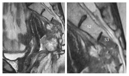
Figure 4 Posterior recurrence involving the presacral fascia (outlined in red).
Figure 5 Posterior recurrence invading into distal sacrum.
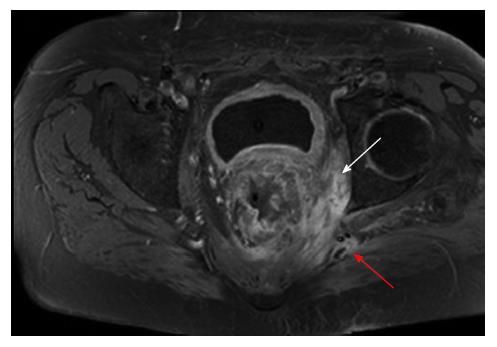
Figure 6 Recurrent rectal cancer in the lower left lateral compartment invading the obturator internus muscle (white arrow) and posteriorly involving the superior gluteal nerve (red arrow).
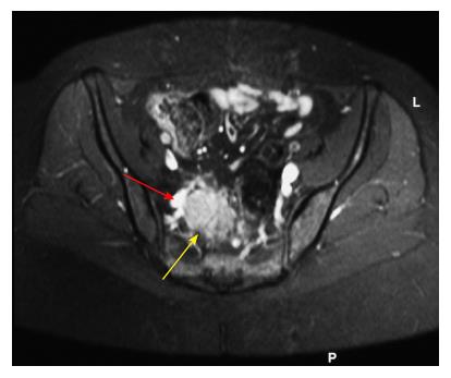
Figure 7 Recurrent rectal cancer at right lateral side wall (yellow arrow), with close proximity to iliac vessels (red arrow); this requires excision of involved vascular segment and reconstruction.
- Citation: Lee DJK, Sagar PM, Sadadcharam G, Tan KY. Advances in surgical management for locally recurrent rectal cancer: How far have we come? World J Gastroenterol 2017; 23(23): 4170-4180
- URL: https://www.wjgnet.com/1007-9327/full/v23/i23/4170.htm
- DOI: https://dx.doi.org/10.3748/wjg.v23.i23.4170


