Copyright
©The Author(s) 2017.
World J Gastroenterol. Jun 14, 2017; 23(22): 4127-4131
Published online Jun 14, 2017. doi: 10.3748/wjg.v23.i22.4127
Published online Jun 14, 2017. doi: 10.3748/wjg.v23.i22.4127
Figure 1 Images obtained during endoscopic examination.
A: There is a slightly raised ulcerated area on the anterior wall of the antrum; B: A positive rolling sign is present in the mid-body, suggestive of a submucosal lesion; C: A diffuse erythematous lesion is seen in the fundic area. These three lesions underwent biopsy sampling.
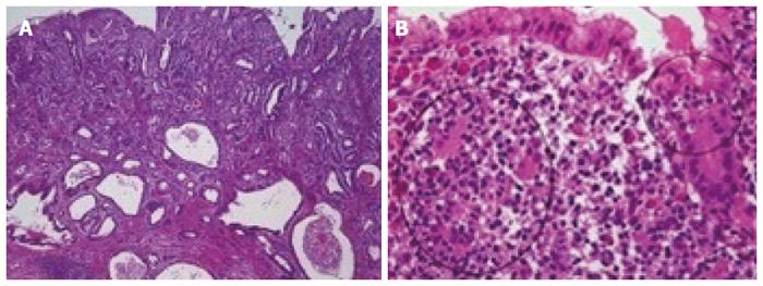
Figure 2 Histologic images.
A: The tumor in the antrum is a moderately differentiated adenocarcinoma [× 40, hematoxylin and eosin (HE)]; B: In the fundus section, lymphoepithelial lesions (circled in black), typical for mucosa-associated lymphoid tissue (MALT) lymphoma, are seen, which are formed by infiltration of centrocyte-like cells into the gastric glandular epithelium (× 400, HE).
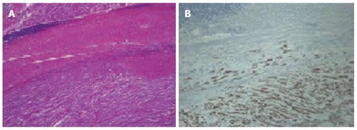
Figure 3 Histologic images.
A: The tumor in the mid-body consists of spindle cells. Above the spindle cell tumor, a smooth muscle layer, lymphoid cuffs, and fundic-type glands are found (× 40, HE); B: On immunohistochemical analysis, the spindle tumor cells are positive for S-100, in contrast to the overlying smooth muscles fibers and lymphoid cells (× 100).
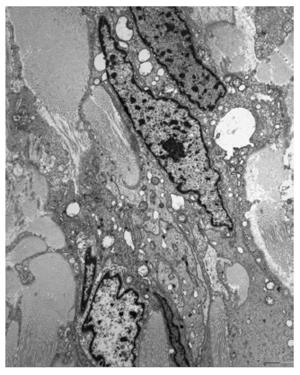
Figure 4 Electron microscopic findings of the submucosal tumor.
Elongated neoplastic cells show with numerous complex interdigitating cytoplasmic processes. The cytoplasmic membrane was completely covered with external lamina. The nuclei reveal irregular margins and a heterochromatic chromatin pattern, and the perikaryal cytoplasm contained several mitochondria, rough endoplasmic reticulum, ribosomes, and many lysosomes.
- Citation: Choi KW, Joo M, Kim HS, Lee WY. Synchronous triple occurrence of MALT lymphoma, schwannoma, and adenocarcinoma of the stomach. World J Gastroenterol 2017; 23(22): 4127-4131
- URL: https://www.wjgnet.com/1007-9327/full/v23/i22/4127.htm
- DOI: https://dx.doi.org/10.3748/wjg.v23.i22.4127


