©The Author(s) 2017.
World J Gastroenterol. May 28, 2017; 23(20): 3702-3712
Published online May 28, 2017. doi: 10.3748/wjg.v23.i20.3702
Published online May 28, 2017. doi: 10.3748/wjg.v23.i20.3702
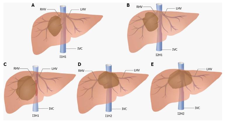
Figure 1 Classifications of liver lesions.
A: Type I1H1; B: I2H1; C: I3H1; D: I1H2; E: I2H2. RHV: Right hepatic vein; LHV: Left hepatic vein; IVC: Inferior vena cava.
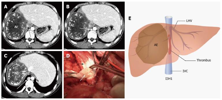
Figure 2 One patient with alveolar echinococcosis in the right lobe of liver.
A: The IVC wall was totally occluded (longer black arrow); B and C: The azygos vein was dilated gradually (longer white arrows); D: The retrohepatic IVC was totally occluded, filled with organized thrombus (shorter white arrow). The shorter black arrow: IVC; E: Classification of this patient was I3H1. LHV: Left hepatic vein; IVC: Inferior vena cava; AE: Alveolar echinococcosis.
- Citation: Li W, Han J, Wu ZP, Wu H. Surgical management of liver diseases invading the hepatocaval confluence based on IH classification: The surgical guideline in our center. World J Gastroenterol 2017; 23(20): 3702-3712
- URL: https://www.wjgnet.com/1007-9327/full/v23/i20/3702.htm
- DOI: https://dx.doi.org/10.3748/wjg.v23.i20.3702


