Copyright
©The Author(s) 2017.
World J Gastroenterol. Mar 28, 2017; 23(12): 2258-2265
Published online Mar 28, 2017. doi: 10.3748/wjg.v23.i12.2258
Published online Mar 28, 2017. doi: 10.3748/wjg.v23.i12.2258
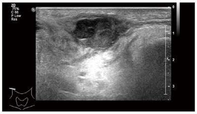
Figure 1 Ultrasonogram of the neck showed a 15 mm × 27 mm mass in the right parotid gland.
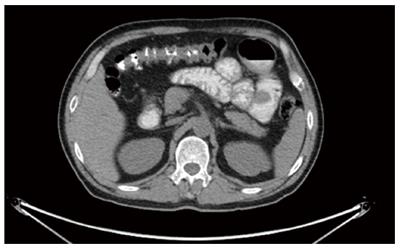
Figure 2 positron emission tomography/computed tomography showed a 36 mm × 33 mm intestinal mass with multiple peripheral lymph nodes in the right midabdomen.
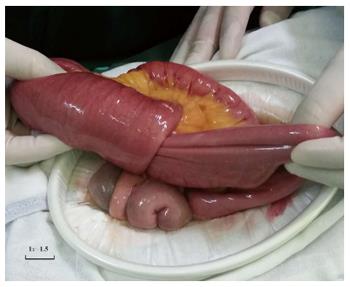
Figure 3 Intussusception was observed 80 cm distal to the duodenojejunal junction and the involved bowels were swollen and expanded.
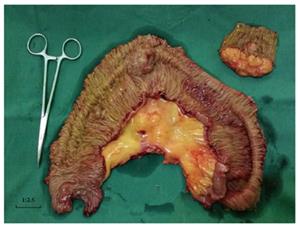
Figure 4 Involved bowels with the masses and mesentery were resected with a proximal 10 cm and distal 10 cm margin.
Figure 5 Microscopic observation of intestinal neoplasms and parotid gland neoplasms.
A: Microphotography shows that polygonal malignant cells of intestinal neoplasms were separated by fibrous tissues, arranging in sheets and nests, with eosinophilic or clear cytoplasm and there was no exact necrosis, vessel invasion and nerve invasion. Nucleolus was obvious and the mitotic index exceeded 20/10 HPF (Hematoxylin-Eosin G × 10); B: Malignant cells of parotid gland neoplasms were similar to the intestinal tumor by microphotography (Hematoxylin-Eosin G × 10).
- Citation: Su H, Liu WS, Ren WH, Wang P, Shi L, Zhou HT. Multiple clear-cell sarcomas of small intestine with parotid gland metastasis: A case report. World J Gastroenterol 2017; 23(12): 2258-2265
- URL: https://www.wjgnet.com/1007-9327/full/v23/i12/2258.htm
- DOI: https://dx.doi.org/10.3748/wjg.v23.i12.2258


