Copyright
©The Author(s) 2017.
World J Gastroenterol. Jan 7, 2017; 23(1): 178-184
Published online Jan 7, 2017. doi: 10.3748/wjg.v23.i1.178
Published online Jan 7, 2017. doi: 10.3748/wjg.v23.i1.178
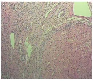
Figure 1 Postoperative pathology.
Nodular cirrhosis associated with hepatocyte and capillary bile cholestasis.
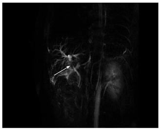
Figure 2 Magnetic resonance cholangiopancreatography findings.
Post- orthotopic liver transplantation anastomotic stenosis of the choledochal duct, intra-hepatic bile duct dilation, and biliary sludge in the common hepatic duct and bilateral hepatic ducts; patient diagnosed with transplantation-related ischemic injury involving the biliary tract.
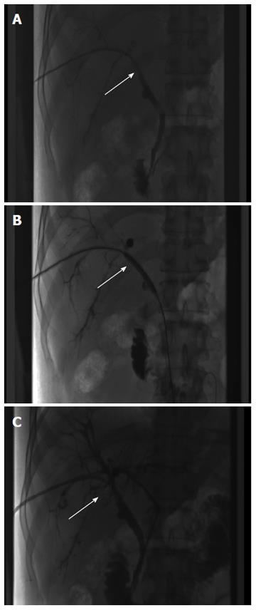
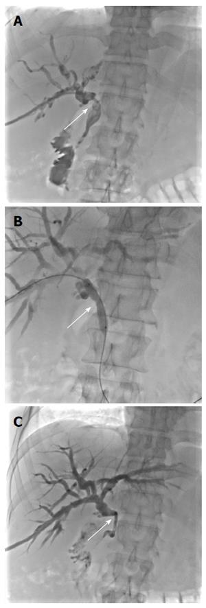
Figure 3 Percutaneous transhepatic cholangial drainage combined with balloon dilation.
A: Anastomotic stenosis of the choledochal duct (straight arrow); B: The inflated balloon (diameter 8 mm, length 4 cm) has a waist at the narrowest part of the stenosis (straight arrow); C: Resolution of the stenosis after balloon dilation (straight arrow).
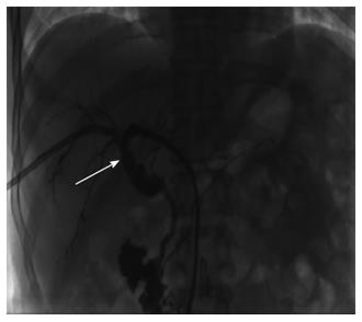
Figure 4 Cholangiography findings.
The anastomotic stenosis was reduced by about 20 % (straight arrow).
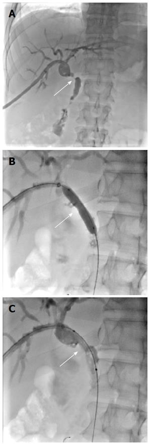
Figure 5 Cutting balloon therapy.
A: Cholangiography showed the development of anastomotic stenosis (straight arrow); B: The inflated cutting balloon (diameter 6 mm, length 4 cm) has a waist at the narrowest part of the stenosis (straight arrow); C: Resolution of the stenosis after balloon dilation (straight arrow).
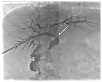
Figure 6 Cholangiography findings.
The anastomotic stenosis was resolved (straight arrow).
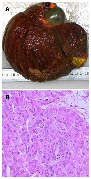
Figure 7 Postoperative pathology.
A: Moderately differentiated hepatocellular carcinoma; B: Peripheral hepatic tissues revealed nodular cirrhosis pathologic changes.
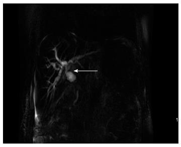
Figure 8 Magnetic resonance cholangiopancreatography findings.
Severe anastomotic stenosis of the choledochal duct, severe choledochectasia involving the intrahepatic bile ducts and left-right hepatic bile ducts above the anastomotic stomas; Patient diagnosed with biliary anastomotic stenosis.
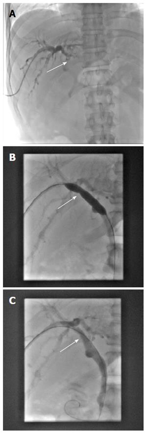
Figure 9 Percutaneous transhepatic cholangial drainage combined with balloon dilation.
A: Severe anastomotic stenosis of the choledochal duct (straight arrow); B: The inflated balloon (diameter 8 mm, length 4 cm) has a waist at the narrowest part of the stenosis (straight arrow); C: Resolution of the stenosis after balloon dilation (straight arrow).
Figure 10 Cutting balloon therapy.
A: Cholangiography showed that the anastomotic stenosis had resolved; B: The inflated cutting balloon (diameter 5 mm, length 2 cm) has a waist at the narrowest part of the stenosis (straight arrow); C: Resolution of the stenosis after balloon dilation (straight arrow).
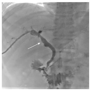
Figure 11 Cholangiography findings.
The anastomotic stenosis was reduced by about 30% (straight arrow).
- Citation: Ding F, Tang H, Xu C, Jiang ZB, Yi SH, Li H, Jiang N, Chen WJ, Yang Q, Yang Y, Chen GH. Cutting balloon treatment of anastomotic biliary stenosis after liver transplantation: Report of two cases. World J Gastroenterol 2017; 23(1): 178-184
- URL: https://www.wjgnet.com/1007-9327/full/v23/i1/178.htm
- DOI: https://dx.doi.org/10.3748/wjg.v23.i1.178


