Copyright
©The Author(s) 2017.
World J Gastroenterol. Jan 7, 2017; 23(1): 167-172
Published online Jan 7, 2017. doi: 10.3748/wjg.v23.i1.167
Published online Jan 7, 2017. doi: 10.3748/wjg.v23.i1.167
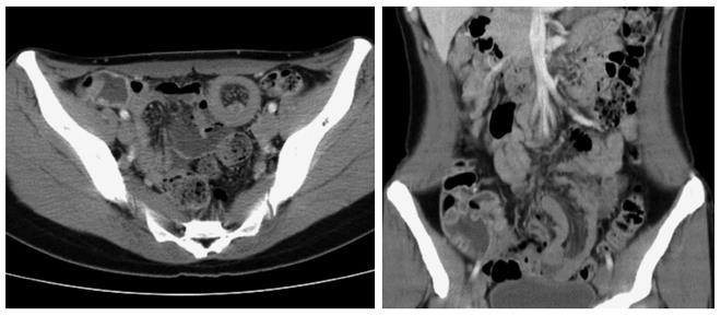
Figure 1 Computed tomography of abdomen: Computed tomography revealed an ileo-ileal intussusception.
The leading point revealed hypovascular mass measuring 1 cm in a diameter.
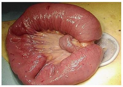
Figure 2 The lesion was externalized using forceps and hands through the umbilical incision.
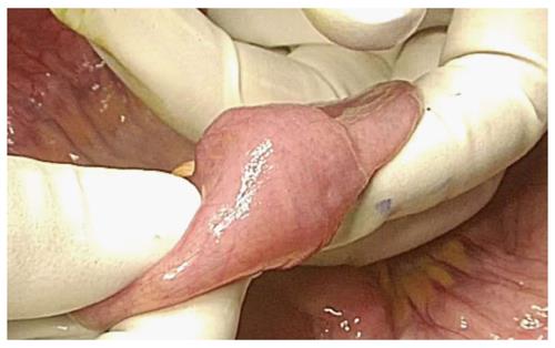
Figure 3 Leading point of intussusception was palpable and soft mass was confirmed.
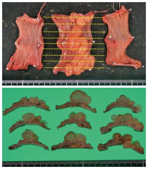
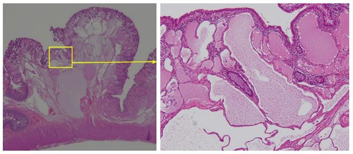
Figure 4 Microscopic findings of the resected specimen.
Polycystic mass with convolutional pattern was found. Cutting lines are shown by lines.
Figure 5 Microscopic findings: Cysts were lined by a flat epithelial endothelium.
No blood cells were found.
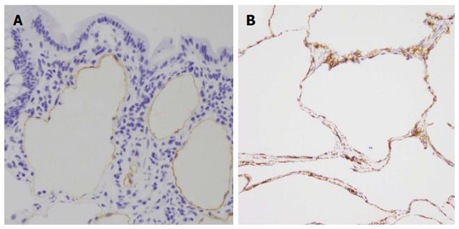
Figure 6 Immunohistochemical examination: The endothelial cells were partially positive for D2-40 (A) and CD34 (B).
- Citation: Kohga A, Kawabe A, Hasegawa Y, Yajima K, Okumura T, Yamashita K, Isogaki J, Suzuki K, Komiyama A. Ileo-ileal intussusception caused by lymphangioma of the small bowel treated by single-incision laparoscopic-assisted ileal resection. World J Gastroenterol 2017; 23(1): 167-172
- URL: https://www.wjgnet.com/1007-9327/full/v23/i1/167.htm
- DOI: https://dx.doi.org/10.3748/wjg.v23.i1.167


