©The Author(s) 2016.
World J Gastroenterol. Feb 7, 2016; 22(5): 1891-1901
Published online Feb 7, 2016. doi: 10.3748/wjg.v22.i5.1891
Published online Feb 7, 2016. doi: 10.3748/wjg.v22.i5.1891
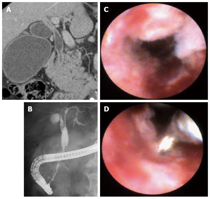
Figure 1 Single-operator cholangiopancreatoscopy procedure.
A: Computed tomography revealed a hilar and inferior bile duct stricture and wall thickening; B: Endoscopic retrograde cholangiogram showed a nonconsecutive bile duct stricture in the hilar and inferior bile ducts; C: SpyGlass view revealed an irregular granular stricture with a thin to thick tortuous vessel; D: Cholangioscope-guided biopsy using the SpyBite was performed in this case.
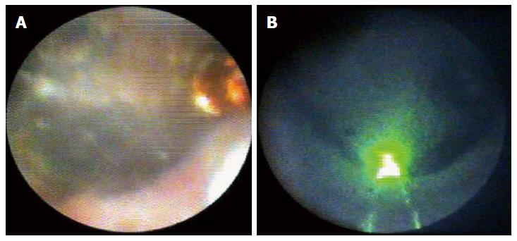
Figure 2 Single-operator cholangiopancreatoscopy view.
A: Obstructive impacted stone targeted by the electrohydraulic lithotripsy probe; B: Obstructive impacted stone targeted by green laser spot.
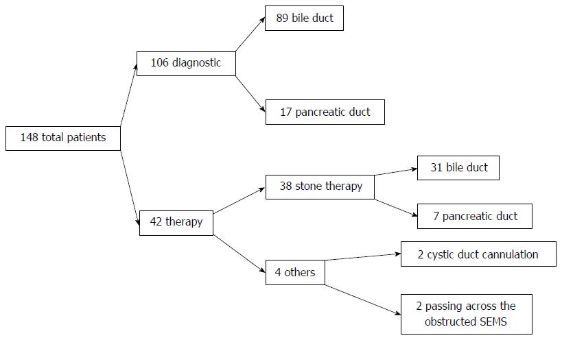
Figure 3 Flow chart of this study.
Overall characteristics of 148 patients who underwent single-operator cholangiopancreatoscopy. SEMS: Self-expandable metal stent.
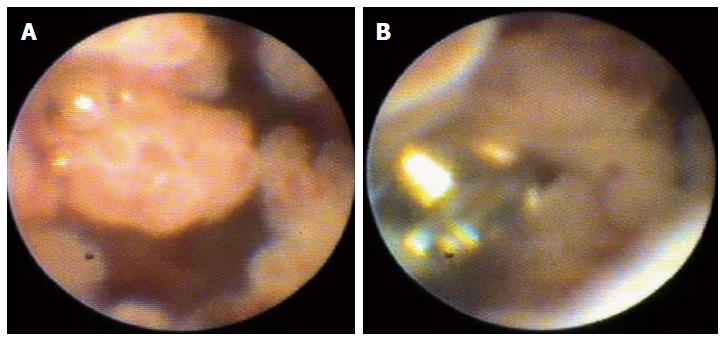
Figure 4 Pancreatoscopy using single-operator cholangiopancreatoscopy for diagnostic purposes.
A: SpyGlass view revealed a papillary and fish-egg-like lesion in the main pancreatic duct; B: Pancreatoscopy-guided biopsy using the SpyBite was performed in this case.
- Citation: Kurihara T, Yasuda I, Isayama H, Tsuyuguchi T, Yamaguchi T, Kawabe K, Okabe Y, Hanada K, Hayashi T, Ohtsuka T, Oana S, Kawakami H, Igarashi Y, Matsumoto K, Tamada K, Ryozawa S, Kawashima H, Okamoto Y, Maetani I, Inoue H, Itoi T. Diagnostic and therapeutic single-operator cholangiopancreatoscopy in biliopancreatic diseases: Prospective multicenter study in Japan. World J Gastroenterol 2016; 22(5): 1891-1901
- URL: https://www.wjgnet.com/1007-9327/full/v22/i5/1891.htm
- DOI: https://dx.doi.org/10.3748/wjg.v22.i5.1891


