Copyright
©The Author(s) 2016.
World J Gastroenterol. Dec 21, 2016; 22(47): 10424-10431
Published online Dec 21, 2016. doi: 10.3748/wjg.v22.i47.10424
Published online Dec 21, 2016. doi: 10.3748/wjg.v22.i47.10424
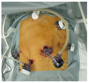
Figure 1 Trocar arrangement for duodenal laparoscopic and endoscopic co-operative surgery.
A 12-mm trocar was placed through the umbilicus as a camera port. Four additional trocars were located symmetrically along the line connecting the location of the target tumor and the umbilicus.
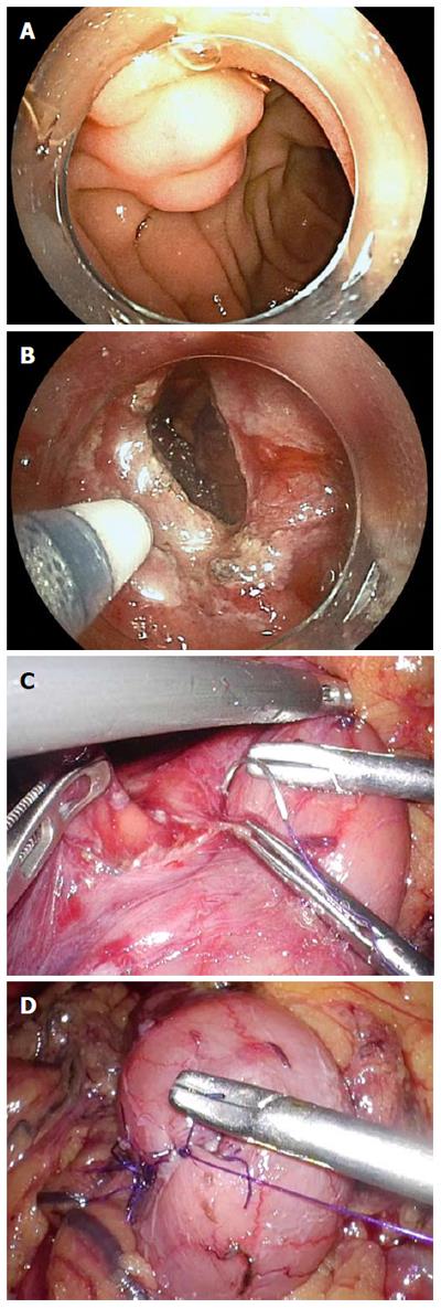
Figure 2 Laparoscopic and endoscopic co-operative surgery procedure for a duodenal submucosal tumors.
A: Endoscopic view of a duodenal neuroendocrine tumor; B: Endoscopic view of a full-thickness dissection; C: Laparoscopic view after full-thickness excision; D: Laparoscopic view after closure using hand-sewn sutures.
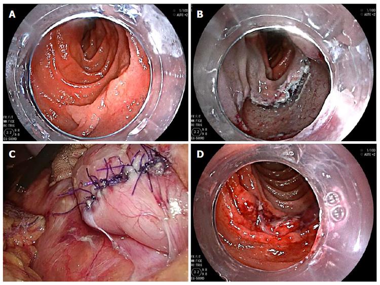
Figure 3 Laparoscopic and endoscopic co-operative surgery procedure for a superficial epithelial tumor.
A: Endoscopic view of a duodenal mucosal tumor; B: Endoscopic view of the ulcer bed after endoscopic submucosal dissection; C: Laparoscopic view after reinforcement using hand-sewn sutures; D: Endoscopic view after the sutures.
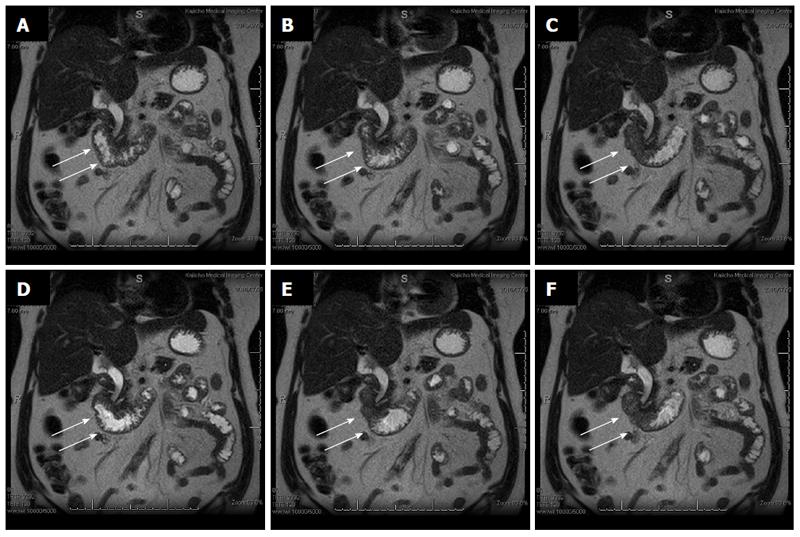
Figure 4 Cine-magnetic resonance imaging findings of duodenal contraction.
These figures are in the following order: A→B→C→D→E→F. White arrows show the second part of the duodenum. They show the presence of periodic active peristalsis in the duodenum.
- Citation: Ichikawa D, Komatsu S, Dohi O, Naito Y, Kosuga T, Kamada K, Okamoto K, Itoh Y, Otsuji E. Laparoscopic and endoscopic co-operative surgery for non-ampullary duodenal tumors. World J Gastroenterol 2016; 22(47): 10424-10431
- URL: https://www.wjgnet.com/1007-9327/full/v22/i47/10424.htm
- DOI: https://dx.doi.org/10.3748/wjg.v22.i47.10424


