©The Author(s) 2016.
World J Gastroenterol. Dec 14, 2016; 22(46): 10254-10259
Published online Dec 14, 2016. doi: 10.3748/wjg.v22.i46.10254
Published online Dec 14, 2016. doi: 10.3748/wjg.v22.i46.10254
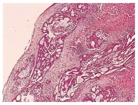
Figure 1 Histologic examination indicated gallbladder cancer with hepatic infiltration.
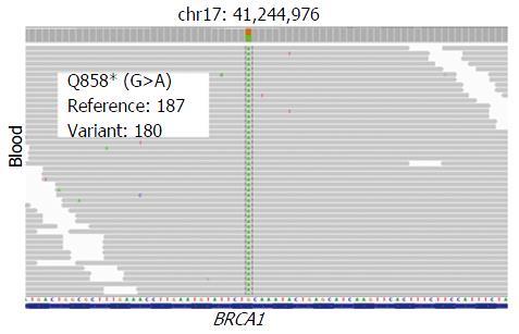
Figure 2 Genomic images from the integrated genome viewer for the alteration in BRCA1 found in the patient’s blood sample.
The number of reads for the reference allele and variant allele are shown for each alteration.
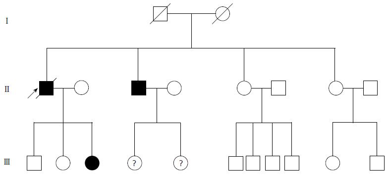
Figure 3 Pedigree of 74-year-old man affected by gallbladder cancer found to be carrier of BRCA1 gene mutation (indicated with arrow).
Black denotes carrier of BRCA1 mutation.
Figure 4 Baseline (July 21, 2015) computed tomography of the abdomen revealed many intra- and extra-hepatic lesions before initiating olaparib treatment.
Figure 5 One month post-olaparib treatment (August 23, 2015).
Computed tomography of the abdomen revealed shrinkage in both the intra- and extra-hepatic lesions and extra-hepatic lesions even appeared to be invisible.
Figure 6 Two and half months post-olaparib treatment (October 9, 2015).
Computed tomography of the abdomen indicated that intrahepatic lesions dwindled; nevertheless, extrahepatic lesions became large and progressed.
- Citation: Xie Y, Jiang Y, Yang XB, Wang AQ, Zheng YC, Wan XS, Sang XT, Wang K, Zhang DD, Xu JJ, Li FG, Zhao HT. Response of BRCA1-mutated gallbladder cancer to olaparib: A case report. World J Gastroenterol 2016; 22(46): 10254-10259
- URL: https://www.wjgnet.com/1007-9327/full/v22/i46/10254.htm
- DOI: https://dx.doi.org/10.3748/wjg.v22.i46.10254


