Copyright
©The Author(s) 2016.
World J Gastroenterol. Nov 21, 2016; 22(43): 9477-9487
Published online Nov 21, 2016. doi: 10.3748/wjg.v22.i43.9477
Published online Nov 21, 2016. doi: 10.3748/wjg.v22.i43.9477
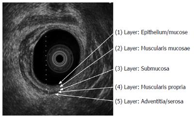
Figure 1 Radial rectal endoscopic ultrasound image: On the right side of the rectum, layering of the rectal wall is retained (white arrows); but normal wall structure has disappeared on the left part of rectum, rectal cancer which involves the third/submucosal layer (uT2).
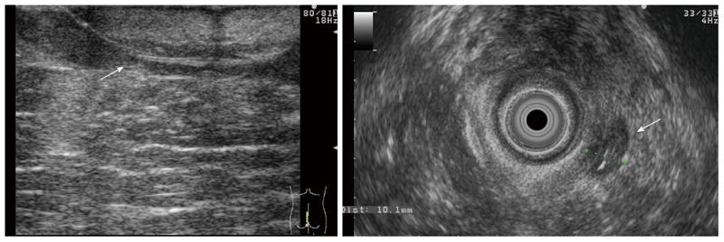
Figure 2 Transabdominal ultrasound image of patient.
A: Active extensive ulcerative colitis. The wall of the transverse colon is widened (largest diameter: 12 mm) and the lumen is narrowed; B: Stricturing ileocolonic Crohn’s disease. Thirty-three millimeter long fibrotic stenosis at the end of the terminal ileum.
Figure 3 Transperineal and rectal ultrasound images of a patient with complicated perianal Crohn’s disease: wide, hypoechoic fistula with seton thread.
Figure 4 Multiplex, hypoechoic pararectal abscesses in rectal and perianal ultrasound images of a patient with perianal Crohn’s disease.
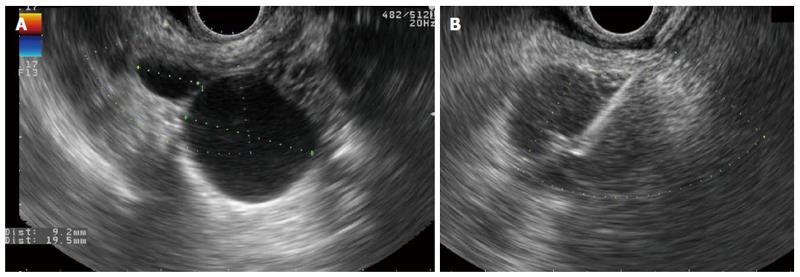
Figure 5 Rectal ultrasound image.
A: Cystic lesion between the rectum and the uterus, it shows typical morphology of endometriosis; B: Inhomogeneous perirectal tissue with rectal wall enlargement and lymphadenomegaly 2-years after resection of rectal cancer. RUS-FNA confirmed the recurrence of rectal adenocarcinoma.
- Citation: Bor R, Fábián A, Szepes Z. Role of ultrasound in colorectal diseases. World J Gastroenterol 2016; 22(43): 9477-9487
- URL: https://www.wjgnet.com/1007-9327/full/v22/i43/9477.htm
- DOI: https://dx.doi.org/10.3748/wjg.v22.i43.9477


