Copyright
©The Author(s) 2016.
World J Gastroenterol. Oct 28, 2016; 22(40): 8883-8891
Published online Oct 28, 2016. doi: 10.3748/wjg.v22.i40.8883
Published online Oct 28, 2016. doi: 10.3748/wjg.v22.i40.8883
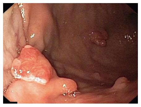
Figure 1 Endoscopic view.
Large gastric hyperplastic polyp.
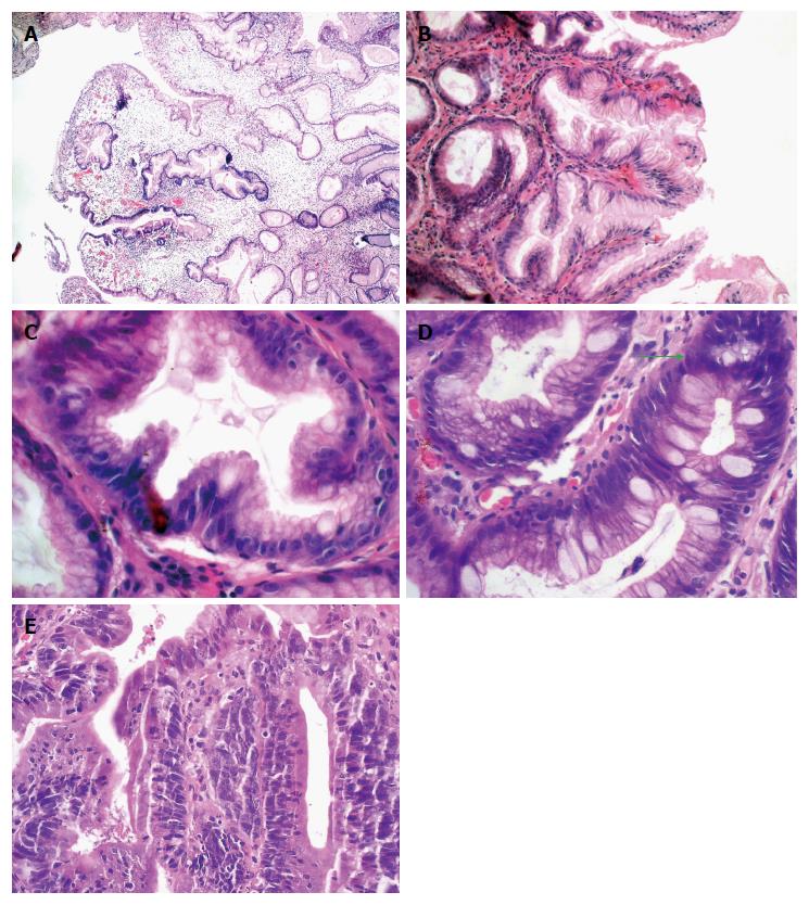
Figure 2 Histopathological findings.
A: Gastric hyperplastic polyp with dilated, elongated, branched and foveolar epithelium and edematous end inflamed stroma (original magnification × 10); B: Gastric hyperplastic polyp with well visible elongated foveolar epthelium (original magnification × 20); C: A cross-section of mucosal crypt shows a serrated ligh of the gland and the goblet cells (original magnification × 40); D: The green arrow indicates a regeneration zone in the foveolar epthelium with hyperchromatic nuclei (original magnification × 40); E: Focus of adenocarcinoma in the gastric hyperplastic polyp (original magnification × 40). Hematoxylin-eosin staining.
- Citation: Markowski AR, Markowska A, Guzinska-Ustymowicz K. Pathophysiological and clinical aspects of gastric hyperplastic polyps. World J Gastroenterol 2016; 22(40): 8883-8891
- URL: https://www.wjgnet.com/1007-9327/full/v22/i40/8883.htm
- DOI: https://dx.doi.org/10.3748/wjg.v22.i40.8883


