©The Author(s) 2016.
World J Gastroenterol. Jan 28, 2016; 22(4): 1593-1606
Published online Jan 28, 2016. doi: 10.3748/wjg.v22.i4.1593
Published online Jan 28, 2016. doi: 10.3748/wjg.v22.i4.1593
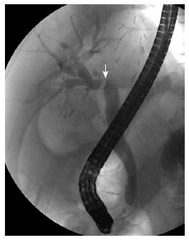
Figure 1 Anastomotic stricture after living donor liver transplantation.
Endoscopic retrograde cholangiography shows a single, tight stricture located at the site of the biliary anastomosis (arrow).
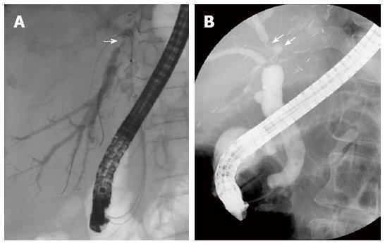
Figure 2 Non-anastomotic strictures after living donor liver transplantation.
Endoscopic retrograde cholangiography shows A biliary stricture at distal right posterior sectoral duct above the anastomotic site (A; arrow); A biliary stricture and dilatation at distal right anterior sectoral duct above the anastomotic site (B; arrows).
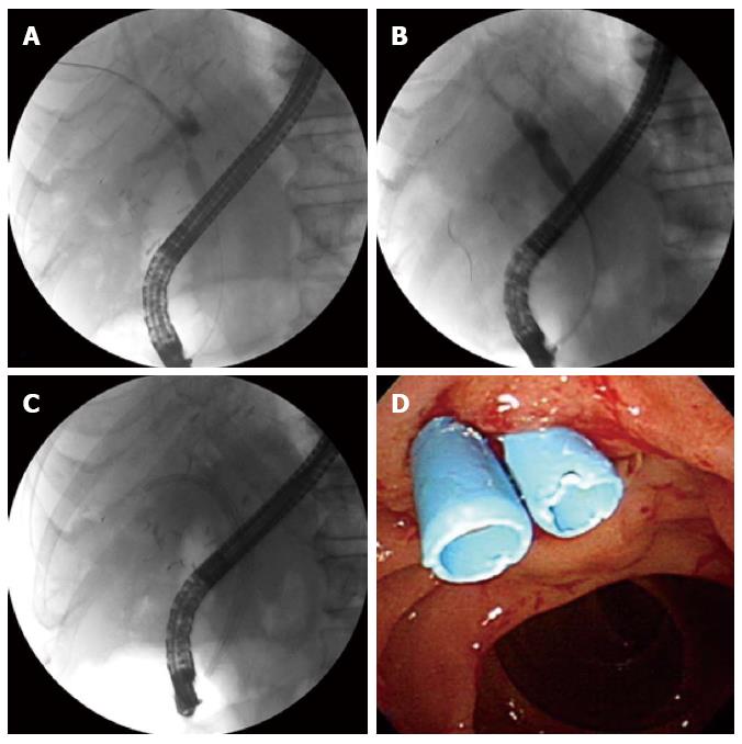
Figure 3 Endoscopic treatment for anastomotic stricture.
Endoscopic retrograde cholangiogram shows an anastomotic stricture (A); balloon dilation of the stricture (B); Two 7 Fr plastic stents are placed across the stricture (C); Endoscopy shows the distal ends of two plastic stents (D).
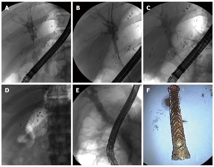
Figure 4 Full-covered self-expanding metallic stent.
Endoscopic retrograde cholangiogram shows an anastomotic stricture (A); Balloon dilation of the stricture (B); A metal stent is placed across the stricture (C); The fully dilated metal stent is shown nine months after placement (D); The anastomotic stricture remains in a fully dilated state after removal of the metal stent (E); A removed metal stent (F).
- Citation: Chang JH, Lee I, Choi MG, Han SW. Current diagnosis and treatment of benign biliary strictures after living donor liver transplantation. World J Gastroenterol 2016; 22(4): 1593-1606
- URL: https://www.wjgnet.com/1007-9327/full/v22/i4/1593.htm
- DOI: https://dx.doi.org/10.3748/wjg.v22.i4.1593


