Copyright
©The Author(s) 2016.
World J Gastroenterol. Jan 21, 2016; 22(3): 917-932
Published online Jan 21, 2016. doi: 10.3748/wjg.v22.i3.917
Published online Jan 21, 2016. doi: 10.3748/wjg.v22.i3.917
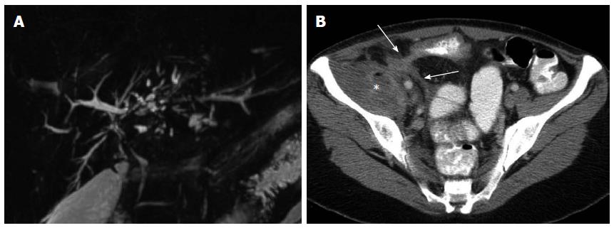
Figure 1 Extraintestinal and extraluminal manifestations of inflammatory bowel disease depicted on imaging.
A: Primary sclerosing cholangitis depicted on magnetic resonance cholangio-pancreatography evidenced as intermittent beading and structuring of the intrahepatic bile ducts; B: Penetrating Crohn’s disease depicted on computed tomography as enhancing fistulous tracks (arrows) of an enterocolonic fistula, with associated abscess formation (asterisk) in the adjacent iliacus musculature.
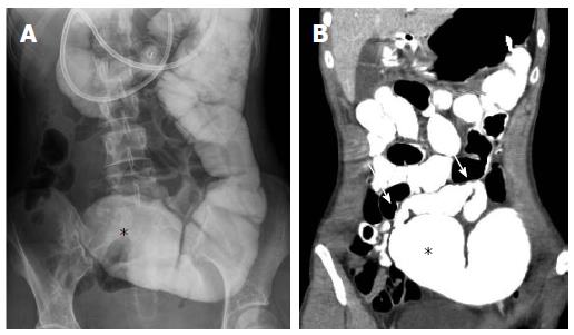
Figure 2 Computed tomography enteroclysis depiction of small bowel stricture.
Fluoroscopic (A) and computed tomography (B) images from an enteroclysis demonstrating a 9 cm stricture in the mid small bowel (arrows) with proximal small bowel dilation (asterisks), which was confirmed during subsequent surgical resection.
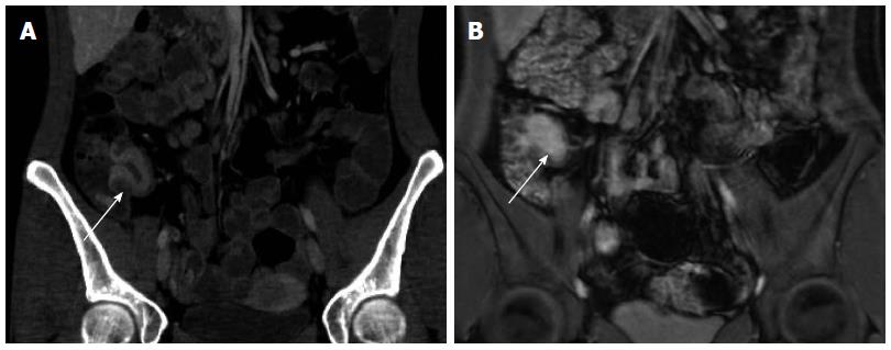
Figure 3 Active Crohn’s terminal ileitis depicted on computed tomography enterography and magnetic resonance enterography in the same patient.
Serial computed tomography enterography (A) and magnetic resonance enterography (B) studies demonstrate marked wall thickening and hyperenhancement (arrows) just proximal to the ileocecal valve consistent with active disease, as confirmed by endoscopy.
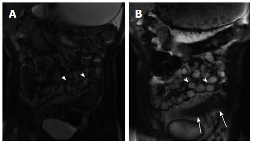
Figure 4 Abnormal bowel peristalsis visualized by cinematic magnetic resonance enterography imaging.
Two static images from a cinematic steady state free precession image series demonstrate multiple normally peristalsing small bowel loops (A, B: arrowheads) as well as a fixed hypoperistaltic loop of inflamed small bowel (B; arrows). This loop also demonstrates wall thickening and mesenteric hypervascularity consistent with active inflammation.
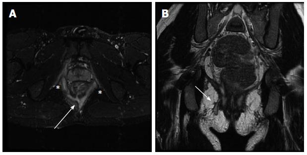
Figure 5 Perianal fistulizing Crohn’s disease depicted on magnetic resonance fistulography.
Axial short tau inversion recovery (A) and coronal (B) T2-weighted (B) images demonstrate a complex perianal fistula at the 6 o’clock position (A; arrow) with an intersphincteric component (A; asterisks) as well as a transphincteric track extending to the skin to the right of midline (B; arrow).
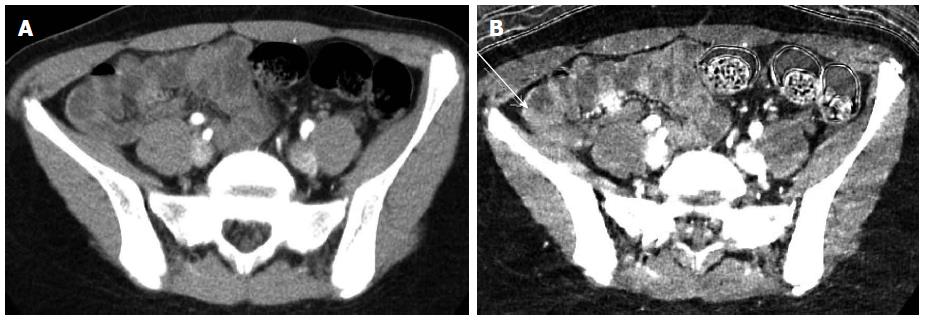
Figure 6 Dual energy computed tomography enterography in inflammatory bowel disease.
Standard (A) and Iodine monochromatic (B) images from a dual energy computed tomography enterography demonstrate a polypoid lesion in the distal ileum (arrow) that was only appreciated on the Iodine images and found to represent an inflammatory pseudopolyp at endoscopy.
- Citation: Kilcoyne A, Kaplan JL, Gee MS. Inflammatory bowel disease imaging: Current practice and future directions. World J Gastroenterol 2016; 22(3): 917-932
- URL: https://www.wjgnet.com/1007-9327/full/v22/i3/917.htm
- DOI: https://dx.doi.org/10.3748/wjg.v22.i3.917


