©The Author(s) 2016.
World J Gastroenterol. May 14, 2016; 22(18): 4501-4514
Published online May 14, 2016. doi: 10.3748/wjg.v22.i18.4501
Published online May 14, 2016. doi: 10.3748/wjg.v22.i18.4501
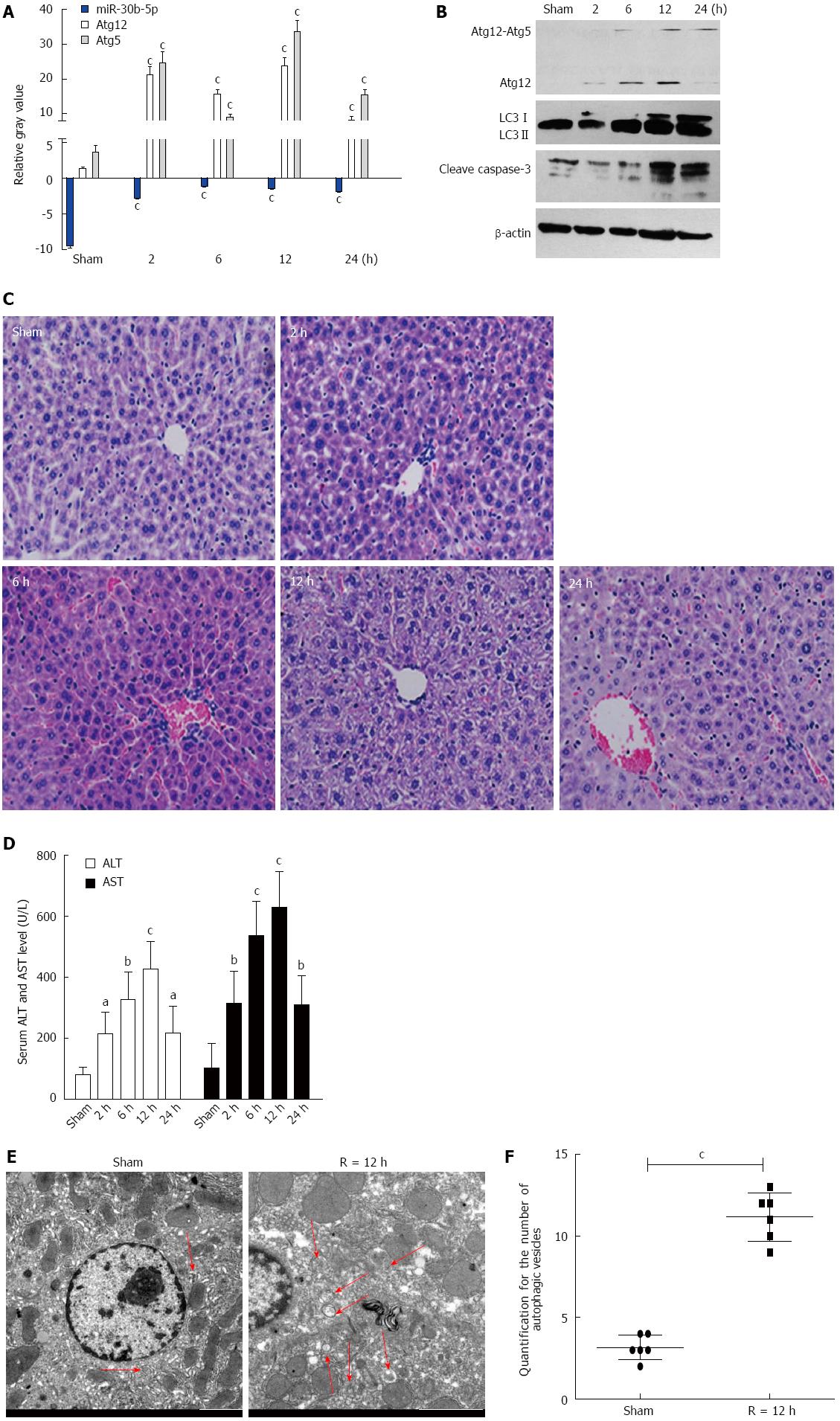
Figure 1 Alterations of miR-30b and autophagy in mouse livers in response to ischemia-reperfusion injury.
A: The expression of miR-30b in mouse livers subjected to IR as determined by quantitative real time polymerase chain reaction analysis; B: Western blotting for autophagy-related gene (Atg) 12, light chain 3 (LC3), and cleave caspase-3 in mouse liver; C: IR treatment increases the histopathologic changes in liver (magnification × 200); D: IR treatment increased serum alanine aminotransferase (ALT) and aspartate aminotransferase (AST) levels in mice compared with sham; E: Transmission electron microscopy (TEM) images of mouse hepatocytes after ischemia followed by reperfusion at 12 h. Scale bars = 2.0 μm. The data were quantified by counting the number of autophagosomes per cross-sectioned cell. aP < 0.05,bP < 0.01,cP < 0.001 vs Sham group. Every experiment was repeated three times.
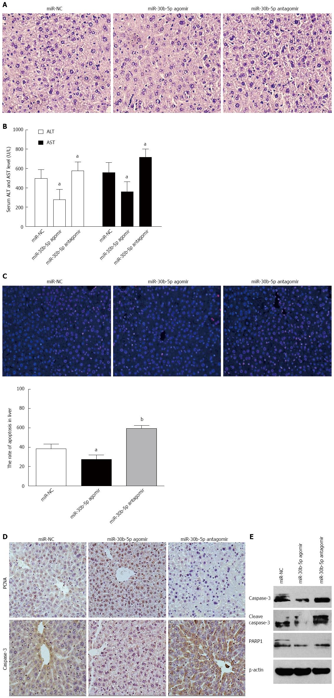
Figure 2 miR-30b can alleviate mouse hepatic ischemia-reperfusion injury.
A: Hematoxylin and eosin (HE) stain was used to observe histopathologic changes in mouse liver (magnification × 200) after tail intravenous injection of miR-30b-5p agomir or antagomir at 12 h following reperfusion; and B: serum AST and ALT levels; C: Cell apoptosis as measured by terminal uridine nick end labeling (TUNEL). Representative sections as determined at 12 h post-reperfusion (magnification × 200); D: Immunohistochemistry revealed expression of proliferating cell nuclear antigen (PCNA) and caspase-3 (magnification × 200); E: Western blot was used to detect expression of caspase-3, cleave caspase-3, and poly ADP-ribose polymerase 1 (PARP1) in livers, aP < 0.05,bP < 0.01 vs miR-NC group. Every experiment was repeated three times.
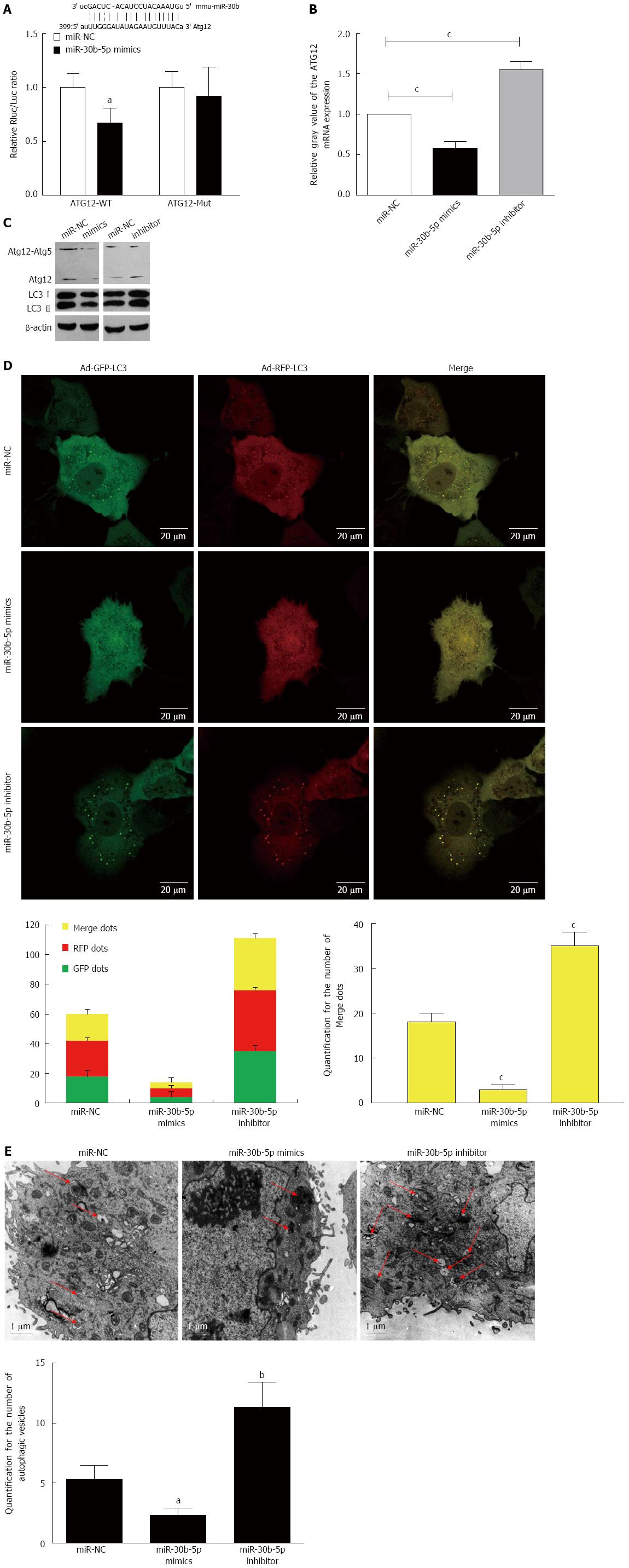
Figure 3 miR-30b inhibits autophagy by sequestering Atg12 in AML12 cells.
A and B: The predicted miR-30b binding site on the Atg12 mRNA 3′-untranslated region (UTR) is shown, and a luciferase reporter assay was performed to determine the effects of miR-30b on the Atg12 mRNA 3′-UTR; C: Western blot was used to detect expression of Atg12 and LC3 in AML12 cells treated with miR-30b mimics or inhibitor; D: Confocal immunofluorescence of AML12 cells demonstrated increased numbers of GFP-RFP-LC3 dots in the IR group; when autophagy is induced, both GFP and RFP are expressed as yellow dots (autophagosomes). Red dots represent autolysosomes as the GFP degrades in an acid environment. Scale bars = 20 μm; E: TEM images of AML12 cells after ischemia followed by reperfusion at 12 h. TEM images show representative examples of autophagosomes (red arrows). Scale bars = 1.0 μm. And the data were quantified by counting the number of autophagosomes per cross-sectioned cell; aP < 0.05, bP < 0.01, cP < 0.001 vs miR-NC group. Every experiment was repeated three times.
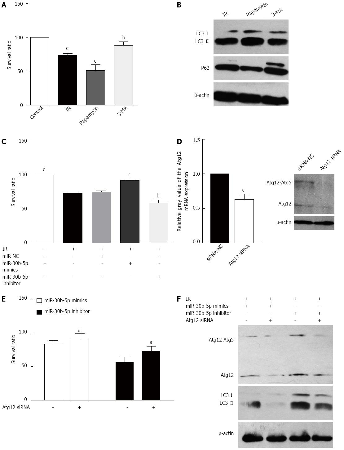
Figure 4 miR-30b alleviate AML12 cell ischemia-reperfusion injury by targeting Atg12 in vitro.
A: The survival ratio of AML12 cells was measured after treatment with rapamycin or 3-MA; B: Western blot was used to detect the expression of LC3 and P62 in AML12 cells treated with Rapamycin or 3-MA; C: The survival ratio of AML12 cells was measured after treatment with miR-30b mimics or inhibitor; D: AML12 cells were transfected with Atg12 siRNAs, and the Atg12 mRNA and protein level of the target were evaluated using quantitative real time polymerase chain reaction or western blot analysis, respectively; E: The survival ratio of AML12 cells was measured at 12 h post-reperfusion in the absence or presence of miR-30b mimics, the miR-30b inhibitor, or Atg12 siRNA; F: Protein levels of Atg12 and LC3 were analyzed using western blot in AML12 cells at 12 h post-reperfusion in the absence or presence of miR-30b mimics, the miR-30b inhibitor, or Atg12 siRNA; aP < 0.05, bP < 0.01, cP < 0.001 vs IR/miR-NC group. Every experiment was repeated three times.
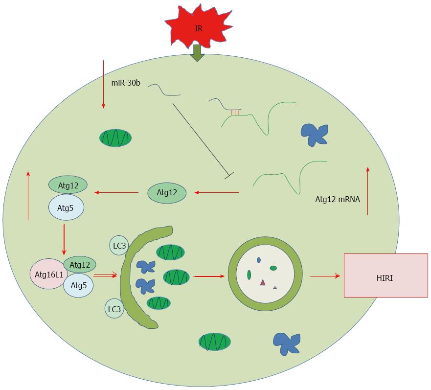
Figure 5 Model of miR-30b inhibition of autophagy to alleviate hepatic ischemia-reperfusion injury via decreasing the Atg12-Atg5 conjugate.
The expression of miR-30b, which binds the 3’-UTR of Atg12, was significantly down-regulated after HIRI. Levels of Atg12 and Atg12-Atg5 conjugate then increased and promoted autophagy, leading to apoptotic cell death. Therefore, miR-30b can inhibit autophagy to alleviate hepatic IRI by decreasing levels of Atg12-Atg5 conjugate. HIRI: Hepatic ischemia-reperfusion injury.
- Citation: Li SP, He JD, Wang Z, Yu Y, Fu SY, Zhang HM, Zhang JJ, Shen ZY. miR-30b inhibits autophagy to alleviate hepatic ischemia-reperfusion injury via decreasing the Atg12-Atg5 conjugate. World J Gastroenterol 2016; 22(18): 4501-4514
- URL: https://www.wjgnet.com/1007-9327/full/v22/i18/4501.htm
- DOI: https://dx.doi.org/10.3748/wjg.v22.i18.4501


