Copyright
©The Author(s) 2016.
World J Gastroenterol. Apr 7, 2016; 22(13): 3693-3700
Published online Apr 7, 2016. doi: 10.3748/wjg.v22.i13.3693
Published online Apr 7, 2016. doi: 10.3748/wjg.v22.i13.3693
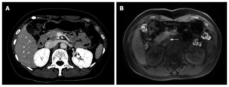
Figure 1 Abdomen computed tomography-scan.
A: CT delayed phase: a relatively low density of nodules (arrow) of approximately 1 cm × 1.4 cm in the uncus of the pancreas; B: Round abnormal signal (arrow) of 1.7 cm × 1.4 cm was found in the head and uncus of the pancreas, T1WI showed low signal, clearly contrasting with normal pancreatic tissue surrounding a relatively high signal.
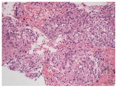
Figure 2 Cytology results.
A: Cells were irregular lumps distributed with medium nuclei sizes, arranged in a disorderly manner, and with crowded overlap; B: Tumor cell with abundant cytoplasm and unclear boundary. Messily arranged spindle cell nuclei can be seen in some cell clumps. Scattered single cells were found in the background.
Figure 3 Biopsy specimen.
Hematoxylin-eosin staining shows the epithelial tumor cells with bright or slightly eosinophilic granules with nested distribution.
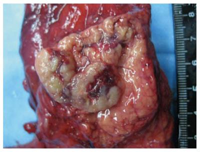
Figure 4 Gross examination.
Tumor 2 cm × 2 cm in size in pancreatic head with clear boundaries. Cross-section of gray and solid soft texture. Metastatic lesions were not found.
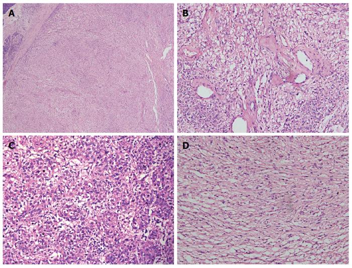
Figure 5 Microscopic examination.
A: Microscopy showing clear boundaries between the tumor and adjacent pancreatic tissue; B: Mounts of vessel were in mesenchyma, with hyaline degeneration present; C: Epithelioid tumor cell; D: Spindle-shaped tumor cell with bright or slightly eosinophilic granule. A, B, C and D: Hematoxylin-eosin staining.
Figure 6 Immunohistochemistry.
A: Tumor cells expressed HMB-45; B: Tumor cells are immunophenotypically positive for the melanocytic marker melan-A; C: Tumor cells are immunophenotypically positive for the smooth muscle marker SMA. Original magnification × 200. A, B and C: Immunostaining.
- Citation: Jiang H, Ta N, Huang XY, Zhang MH, Xu JJ, Zheng KL, Jin G, Zheng JM. Pancreatic perivascular epithelioid cell tumor: A case report with clinicopathological features and a literature review. World J Gastroenterol 2016; 22(13): 3693-3700
- URL: https://www.wjgnet.com/1007-9327/full/v22/i13/3693.htm
- DOI: https://dx.doi.org/10.3748/wjg.v22.i13.3693


