Copyright
©The Author(s) 2016.
World J Gastroenterol. Mar 28, 2016; 22(12): 3502-3505
Published online Mar 28, 2016. doi: 10.3748/wjg.v22.i12.3502
Published online Mar 28, 2016. doi: 10.3748/wjg.v22.i12.3502
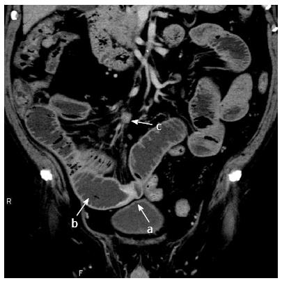
Figure 1 Abdominal computed tomography-scan showed focal thickening of the wall of the small intestine (a), intestinal dilatation of the oral intestine (b), and intraperitoneal lymphadenopathy (c).
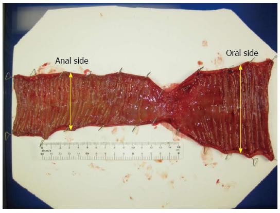
Figure 2 Resected specimen showed a 5-6 cm tumor.
The oral side lumen was 1.5-fold bigger lumen than the anal side.
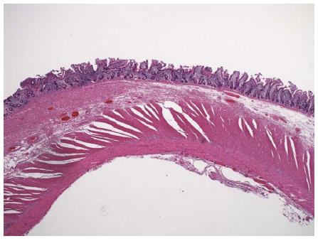
Figure 3 Histologically, we found thickened portions in the wall, muscle plate thickening, fat reduction, fibrosis.
The left half of the specimen is lesion area and the right half is normal area (HE × 10).
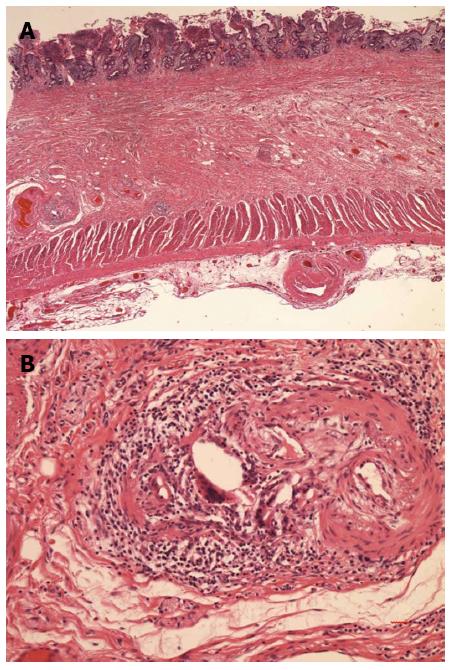
Figure 4 Cholesterol clefts were detected in the thickened portions in the wall.
A: × 20; B: × 200.
- Citation: Azuma S, Ikenouchi M, Akamatsu T, Seta T, Urai S, Uenoyama Y, Yamashita Y. Ileus caused by cholesterol crystal embolization: A case report. World J Gastroenterol 2016; 22(12): 3502-3505
- URL: https://www.wjgnet.com/1007-9327/full/v22/i12/3502.htm
- DOI: https://dx.doi.org/10.3748/wjg.v22.i12.3502


