Copyright
©The Author(s) 2016.
World J Gastroenterol. Mar 28, 2016; 22(12): 3404-3411
Published online Mar 28, 2016. doi: 10.3748/wjg.v22.i12.3404
Published online Mar 28, 2016. doi: 10.3748/wjg.v22.i12.3404
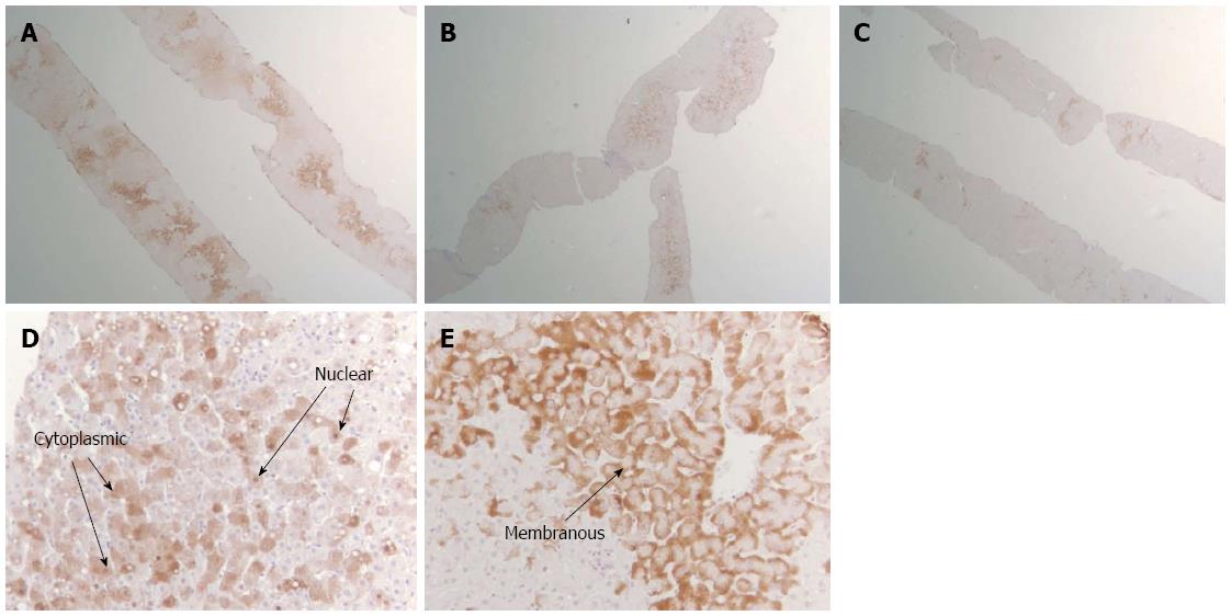
Figure 1 Images of pathology slides illustrating the classification system for intrahepatic hepatitis B surface antigen and hepatitis B core antigen.
A: Diffuse distribution of HBsAg (× 2); B: Patchy distribution of HBcAg (× 2); C: Rare distribution of HBsAg (× 2); D: Cytoplasmic/nuclear pattern of HBcAg (× 20); E: Membranous pattern of HBsAg (× 20).
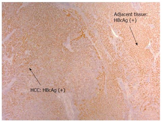
Figure 2 Diffuse hepatitis B core antigen distribution of mixed cytoplasmic, nuclear pattern in both tumor and adjacent non-tumor tissue (× 10).
HCC: Hepatocellular carcinoma; HBcAg: Hepatitis B core antigen.
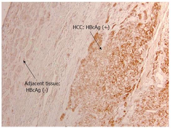
Figure 3 Diffuse hepatitis B core antigen distribution of mixed cytoplasmic, nuclear pattern in tumor tissue alone but not in adjacent non-tumor tissue (× 10).
HCC: Hepatocellular carcinoma; HBcAg: Hepatitis B core antigen.
- Citation: Safaie P, Poongkunran M, Kuang PP, Javaid A, Jacobs C, Pohlmann R, Nasser I, Lau DT. Intrahepatic distribution of hepatitis B virus antigens in patients with and without hepatocellular carcinoma. World J Gastroenterol 2016; 22(12): 3404-3411
- URL: https://www.wjgnet.com/1007-9327/full/v22/i12/3404.htm
- DOI: https://dx.doi.org/10.3748/wjg.v22.i12.3404


