Copyright
©The Author(s) 2015.
World J Gastroenterol. Feb 14, 2015; 21(6): 1982-1988
Published online Feb 14, 2015. doi: 10.3748/wjg.v21.i6.1982
Published online Feb 14, 2015. doi: 10.3748/wjg.v21.i6.1982
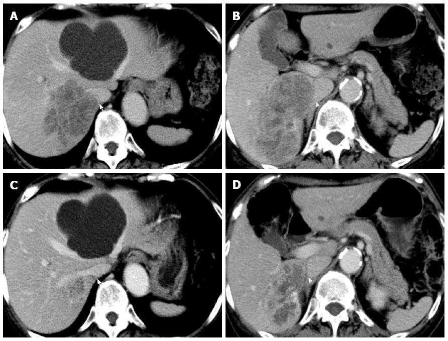
Figure 1 Enhanced computed tomography.
A, B: Before chemotherapy. A huge synchronous colorectal liver metastasis was involving the right hepatic vein (RHV; arrow) and the inferior vena cava (IVC; arrowhead); C, D: After chemotherapy. The tumor was dramatically reduced, and the IVC was isolated from the tumor (arrowhead).
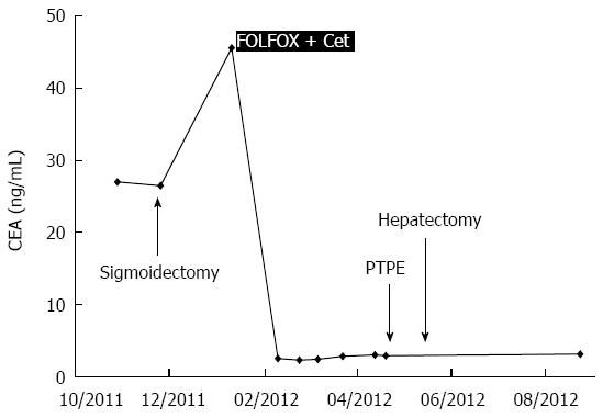
Figure 2 Serum levels of carcinoembryonic antigen.
FOLFOX: 5-fluorouracil, leucovorin, and oxaliplatin; Cet: Cetuximab; PTPE: Percutaneous transhepatic portal vein embolization.
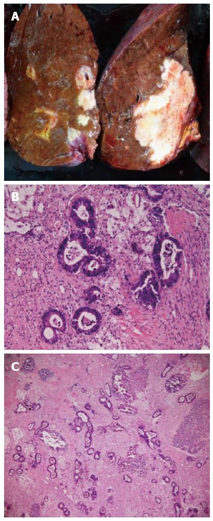
Figure 3 Resected specimen.
A: Cut surface. The tumor was 70 mm × 40 mm in size. The tumor was grayish-white and stony hard; B, C: Hematoxylin and eosin (HE) staining of the resected specimen; B: HE staining, × 400. Adenocarcinoma; C: HE, × 100. Approximately 50% of the adenocarcinoma was necrotic.
- Citation: Baba K, Oshita A, Kohyama M, Inoue S, Kuroo Y, Yamaguchi T, Nakamura H, Sugiyama Y, Tazaki T, Sasaki M, Imamura Y, Daimaru Y, Ohdan H, Nakamitsu A. Successful treatment of conversion chemotherapy for initially unresectable synchronous colorectal liver metastasis. World J Gastroenterol 2015; 21(6): 1982-1988
- URL: https://www.wjgnet.com/1007-9327/full/v21/i6/1982.htm
- DOI: https://dx.doi.org/10.3748/wjg.v21.i6.1982


