Copyright
©The Author(s) 2015.
World J Gastroenterol. Feb 14, 2015; 21(6): 1775-1783
Published online Feb 14, 2015. doi: 10.3748/wjg.v21.i6.1775
Published online Feb 14, 2015. doi: 10.3748/wjg.v21.i6.1775
Figure 1 Serum aspartate aminotransferase and alanine aminotransferase in rats.
A: The serum aspartate aminotransferase (mean ± SE) in rats subjected to 45 min of segmental (left lateral lobe) liver ischemia and 4 h of reperfusion with (nitrite, n = 14) or without (ischemia reperfusion injury, n = 14) intravenous pre-treatment with 480 nmol of nitrite and sham operated animals (n = 8). The animals treated with nitrite prior to ischemia and reperfusion had significantly lower aspartate aminotransferase (AST) levels (P = 0.022); B: The serum alanine aminotransferase (ALT) (mean ± SE) in rats subjected to 45 min of segmental (left lateral lobe) liver ischemia and 4 h of reperfusion with (nitrite, n = 14) or without (ischemia reperfusion injury, n = 14) intravenous pre-treatment with 480 nmol of nitrite and sham operated animals (n = 8). The animals treated with nitrite prior to ischemia and reperfusion had significantly lower AST levels (P = 0.0045). IRI: Ischemia reperfusion injury.
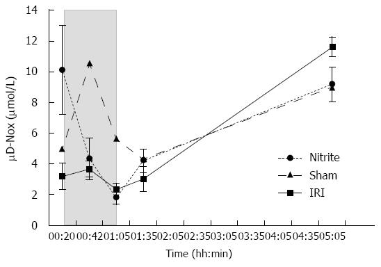
Figure 2 Parenchymal (microdialytic), nitrite and nitrate (mean ± SE) in the ischemic liver lobes in rats subjected to 45 min of segmental (left lateral lobe) liver ischemia and 4 h of reperfusion with (nitrite, n = 14) or without (ischemia reperfusion injury, n = 14) intravenous pre-treatment with 480 nmol of nitrite and sham operated animals (n = 8).
After the administration of nitrite, the parenchymal nitrite and nitrate (NOx) was higher than for animals that did not receive nitrite (P = 0.031). During ischemia, the levels decreased in both groups and then increased again during reperfusion. After 4 h of reperfusion, there was a trend towards higher levels in animals that were not treated with nitrite (P = 0.067). The shaded area represents the ischemic phase. IRI: Ischemia reperfusion injury.
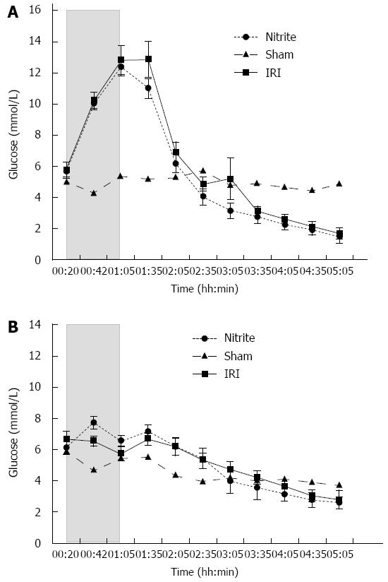
Figure 3 Parenchymal (microdialytic) glucose.
A: Parenchymal (microdialytic) glucose (mean ± SE) in the ischemic liver lobes in rats subjected to 45 min of segmental (left lateral lobe) liver ischemia and 4 h of reperfusion with (nitrite, n = 14) or without (ischemia reperfusion injury, IRI, n = 14) intravenous pre-treatment with 480 nmol of nitrite and sham operated animals (n = 8). During ischemia, parenchymal glucose increased in both groups without any difference noted between the groups. In the reperfusion phase, there was a steady decline in the glucose levels. The shaded area represents the ischemic phase; B: Parenchymal (microdialytic) glucose (mean ± SE) in the control (non-ischemic, right) liver lobes in rats subjected to 45 min of segmental (left lateral lobe) liver ischemia and 4 h of reperfusion with (nitrite, n = 14) or without (IRI, n = 14) intravenous pre-treatment with 480 nmol of nitrite and sham operated animals (n = 8). After the administration of nitrite, parenchymal glucose increased significantly (P < 0.001) and reached levels higher than those in the untreated group (P = 0.02).
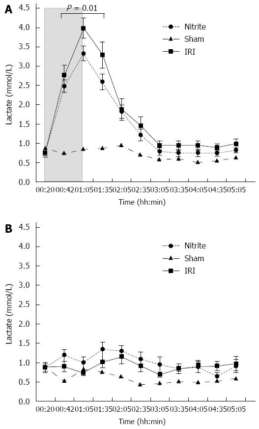
Figure 4 Parenchymal (microdialytic) lactate.
A: Parenchymal (microdialytic) lactate (mean ± SE) in the ischemic liver lobes in rats subjected to 45 min of segmental (left lateral lobe) liver ischemia and 4 h of reperfusion with (nitrite, n = 14) or without (ischemia reperfusion injury, IRI, n = 14) intravenous pre-treatment with 480 nmol of nitrite and sham operated animals (n = 8). During ischemia, parenchymal lactate increased in both groups; during the ischemic phase and first 30 min of the reperfusion phase, the levels were higher in the untreated group (P = 0.01). The shaded area represents the ischemic phase; B: Parenchymal (microdialytic) lactate (mean ± SE) in the control (non-ischemic, right) liver lobes in rats subjected to 45 min of segmental (left lateral lobe) liver ischemia and 4 h of reperfusion with (nitrite, n = 14) or without (IRI, n = 14) intravenous pre-treatment with 480 nmol of nitrite and sham operated animals (n = 8). After the administration of nitrite, the parenchymal lactate increased significantly (P = 0.012) and reached levels higher than those in the untreated group (P = 0.01).
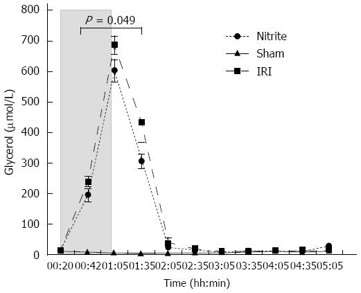
Figure 5 Parenchymal (microdialytic) glycerol (mean ± SE) in the ischemic liver lobes in rats subjected to 45 min of segmental (left lateral lobe) liver ischemia and 4 h of reperfusion with (nitrite, n = 14) or without (ischemia reperfusion injury, n = 14) intravenous pre-treatment with 480 nmol of nitrite and sham operated animals (n = 8).
During ischemia, the parenchymal glycerol increased in both groups; during the ischemic phase and the first 30 min of the reperfusion phase, the levels were higher in the untreated group (P = 0.049). The shaded area represents the ischemic phase. IRI: Ischemia reperfusion injury.
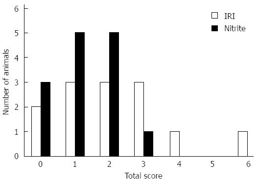
Figure 6 Distribution of total histological injury score.
The distribution of total histological injury score in the ischemic liver lobes in rats subjected to 45 min of segmental (left lateral lobe) liver ischemia and 4 h of reperfusion with (nitrite, n = 14) or without (ischemia reperfusion injury, IRI, n = 14) intravenous pre-treatment with 480 nmol of nitrite.
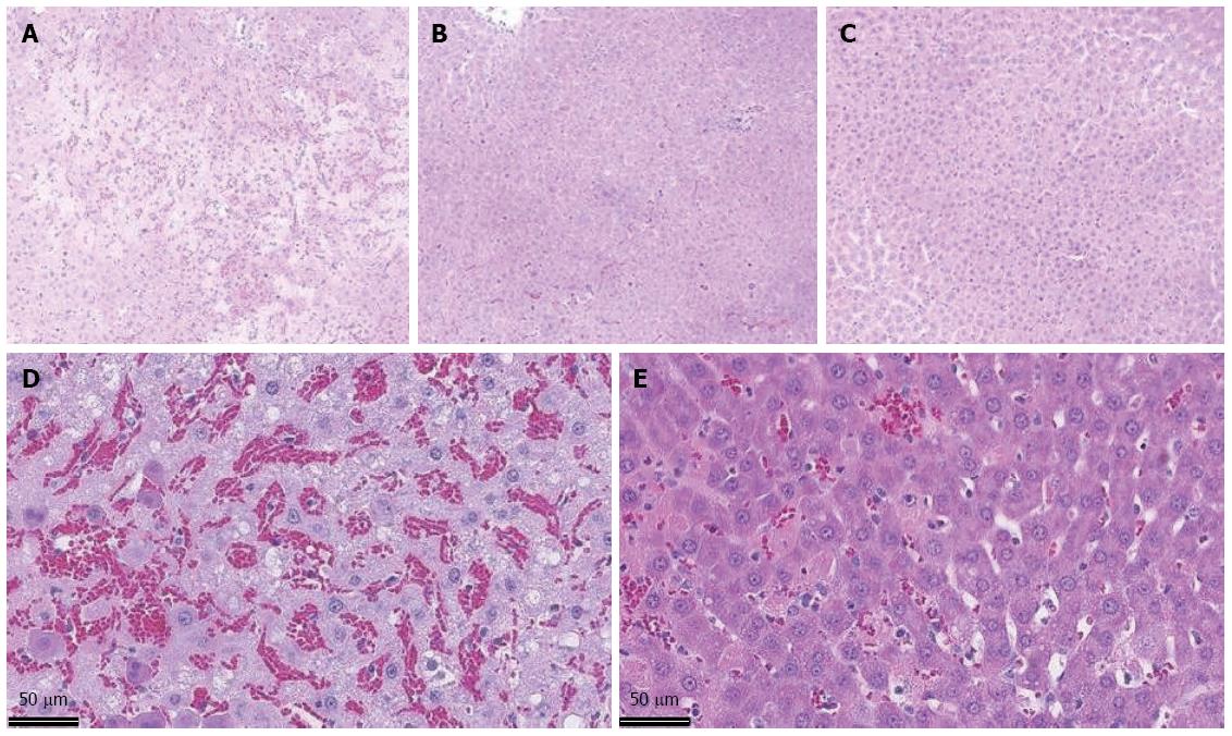
Figure 7 HE-stained liver tissue from the ischemic liver lobes in rats subjected to 45 min of segmental (left lateral lobe) ischemia and 4 h of reperfusion without pre-treatment (A), with 480 nmol of nitrite administered intravenously before ischemia (B) and sham operated (20 x magnification) (C); In 40 × magnificataion, without pre-treatment (D) and with 480 nmol of nitrite administered intravenously before ischemia (E).
- Citation: Björnsson B, Bojmar L, Olsson H, Sundqvist T, Sandström P. Nitrite, a novel method to decrease ischemia/reperfusion injury in the rat liver. World J Gastroenterol 2015; 21(6): 1775-1783
- URL: https://www.wjgnet.com/1007-9327/full/v21/i6/1775.htm
- DOI: https://dx.doi.org/10.3748/wjg.v21.i6.1775


