©The Author(s) 2015.
World J Gastroenterol. Dec 28, 2015; 21(48): 13582-13586
Published online Dec 28, 2015. doi: 10.3748/wjg.v21.i48.13582
Published online Dec 28, 2015. doi: 10.3748/wjg.v21.i48.13582
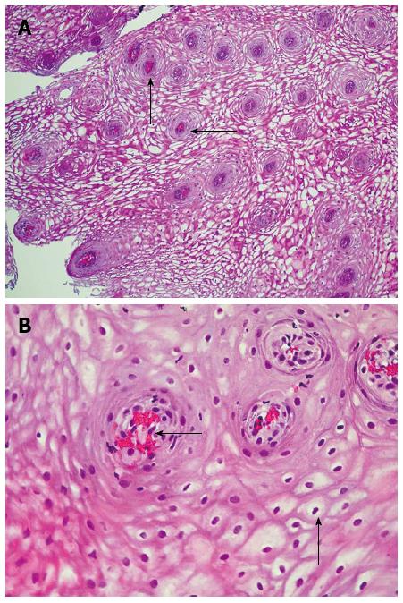
Figure 1 Stratified squamous non-keratinized epithelium stained with haematoxylin and eosin.
A: Focal proliferation of the basal cells (arrow, magnification × 100); B: Plethora of vessels and stasis of erythrocytes in the vessels of lamina propria papillae (arrow), dystrophy of the epithelial cells (arrow, magnification × 400).
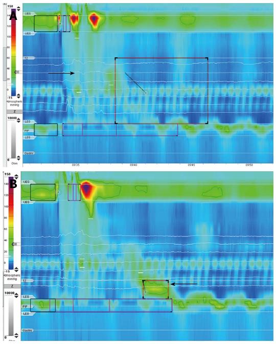
Figure 2 High-resolution manometry.
A: Lack of peristaltic contractions in the thoracic esophagus (arrow); B: The intraesophageal pressure was increased during the passage of the liquid bolus and 1-2 s later in the distal part of the esophagus was registered the spastic contraction (arrow).
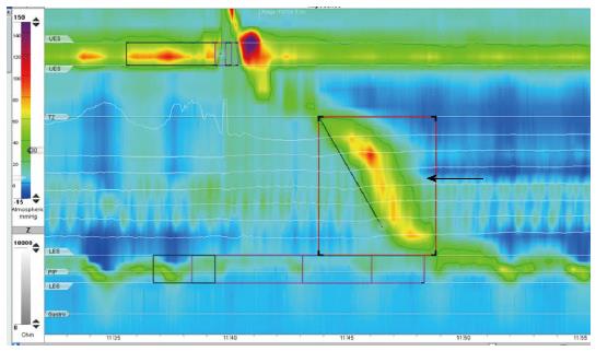
Figure 3 High-resolution manometry (against the background of antithyroid drug therapy) in the thoracic region of the esophagus is noted the normal esophageal peristalsis in response to swallowing of water (arrow).
- Citation: Evsyutina Y, Trukhmanov A, Ivashkin V, Storonova O, Godjello E. Case report of Graves’ disease manifesting with odynophagia and heartburn. World J Gastroenterol 2015; 21(48): 13582-13586
- URL: https://www.wjgnet.com/1007-9327/full/v21/i48/13582.htm
- DOI: https://dx.doi.org/10.3748/wjg.v21.i48.13582


