©The Author(s) 2015.
World J Gastroenterol. Dec 21, 2015; 21(47): 13309-13315
Published online Dec 21, 2015. doi: 10.3748/wjg.v21.i47.13309
Published online Dec 21, 2015. doi: 10.3748/wjg.v21.i47.13309
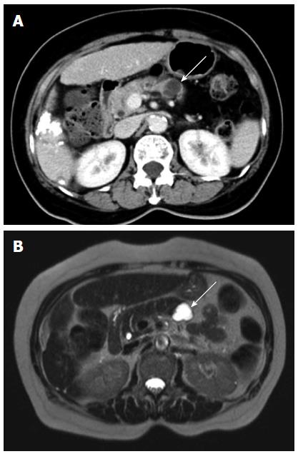
Figure 1 Contrast-enhanced computed tomography scans and T2-weighted magnetic resonance images.
Contrast-enhanced computed tomography scans obtained during the portal venous phase showing a septated cystic lesion in the body of the pancreas (A; arrow). An axial T2-weighted magnetic resonance image showing a T2 hyperintense lesion with septa (B; arrow) in the body of the pancreas.
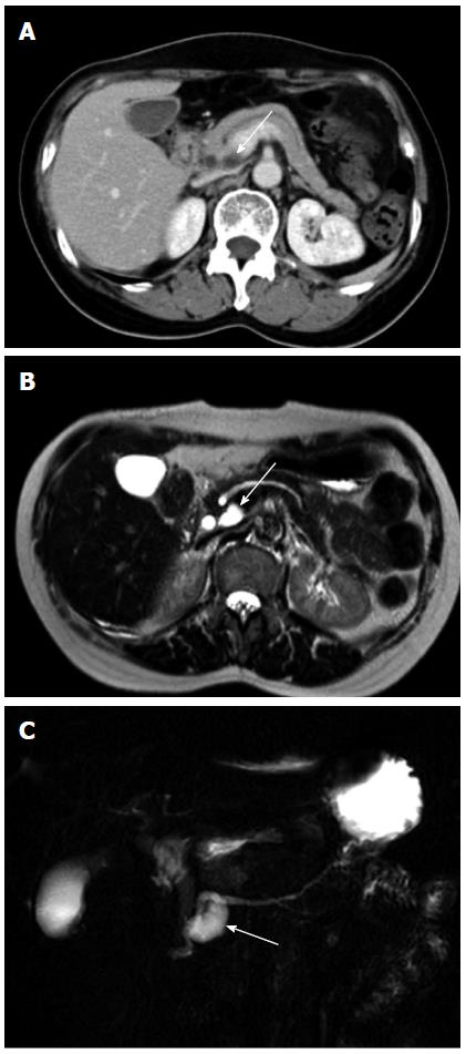
Figure 2 Contrast-enhanced computed tomography scans and T2-weighted magnetic resonance images.
Contrast-enhanced computed tomography scans obtained during the portal venous phase showing a tubular cyst in the neck of the pancreas (A; white arrow). Axial T2-weighted magnetic resonance image showing a T2 hyperintense tubular lesion (B; white arrow) in the neck of the pancreas. The tubular lesion (C; white arrow) was more clearly appreciated on two dimensional magnetic resonance cholangiopancreaticography.
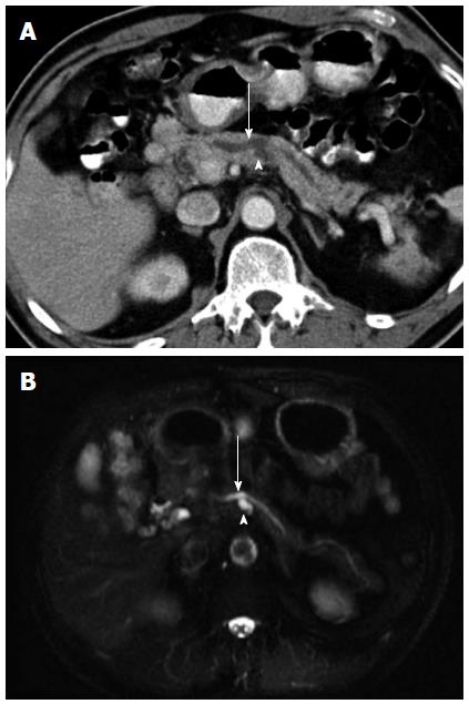
Figure 3 Contrast-enhanced computed tomography scans and T2-weighted magnetic resonance images.
Contrast-enhanced computed tomography scans obtained during the portal venous phase showing a tubular cyst (A; arrowhead) in the body of the pancreas, with a prominent pancreatic duct (A; arrow). Axial T2-weighted magnetic resonance image showing a T2 hyperintense tubular cyst (B; arrowhead) in the body of the pancreas, with a prominent pancreatic duct (B; arrow).
- Citation: Tsai HM, Chuang CH, Shan YS, Liu YS, Chen CY. Features associated with progression of small pancreatic cystic lesions: A retrospective study. World J Gastroenterol 2015; 21(47): 13309-13315
- URL: https://www.wjgnet.com/1007-9327/full/v21/i47/13309.htm
- DOI: https://dx.doi.org/10.3748/wjg.v21.i47.13309


