©The Author(s) 2015.
World J Gastroenterol. Nov 7, 2015; 21(41): 11609-11620
Published online Nov 7, 2015. doi: 10.3748/wjg.v21.i41.11609
Published online Nov 7, 2015. doi: 10.3748/wjg.v21.i41.11609
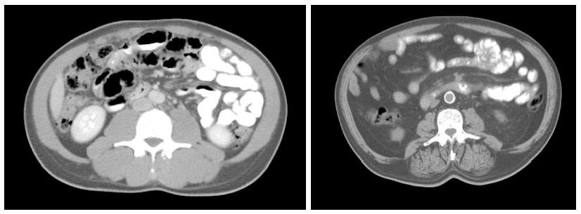
Figure 1 Axial computed tomography images of the third lumbar vertebra region from two patients with similar body mass index but different muscle and fat tissue cross sectional areas.
Paraspinal muscles are clearly different between the two subjects as is mesenteric fat and fat infiltrating muscle - muscle radiation attenuation. Low relative muscularity and expanded visceral fat are associated with increased toxicity and decreased survival.
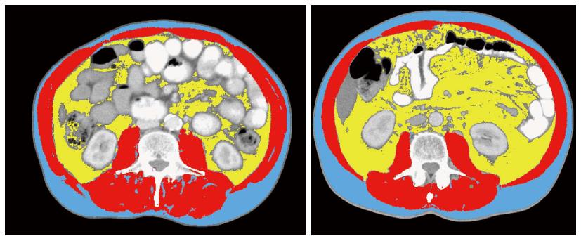
Figure 2 Lumbar computed tomography was analyzed for muscle and fat tissue cross sectional areas using an appropriate software developed by Martin et al[88].
Muscle mass is shown in red and were quantified within a Hounsfield unit (HU) range of -29-150, visceral fat shown in yellow, range from -150 to -50, and subcutaneous fat shown in blue, range from -190 to 30. Muscle radiation attenuation was calculated for muscle area. Although these two images might refer to two individuals with the same body mass index (23 kg/m2) and age (73 yr), the amount of muscle mass and visceral fat, which amplifies inflammatory response, are very distinct.
- Citation: Cravo M, Fidalgo C, Garrido R, Rodrigues T, Luz G, Palmela C, Santos M, Lopes F, Maio R. Towards curative therapy in gastric cancer: Faraway, so close! World J Gastroenterol 2015; 21(41): 11609-11620
- URL: https://www.wjgnet.com/1007-9327/full/v21/i41/11609.htm
- DOI: https://dx.doi.org/10.3748/wjg.v21.i41.11609


