©The Author(s) 2015.
World J Gastroenterol. Aug 21, 2015; 21(31): 9437-9441
Published online Aug 21, 2015. doi: 10.3748/wjg.v21.i31.9437
Published online Aug 21, 2015. doi: 10.3748/wjg.v21.i31.9437
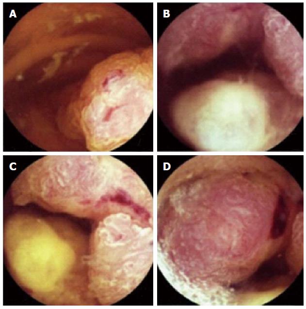
Figure 1 Capsule endoscopy findings in the 3rd and 4th portions of the duodenum.
A: Polypoid and multilobular lesion; B, C: Partially obstructing the lumen; C, D: Ulcerated with low-flow bleeding.
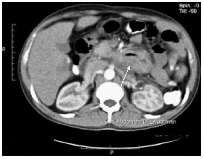
Figure 2 Abdominal computed tomography scan demonstrating a thickening of the wall involving the 3rd and 4th portions of duodenum, narrowing its lumen (arrows), without clear lines of cleavage with adjacent structures and with no evidence of nodal and distant metastasis.
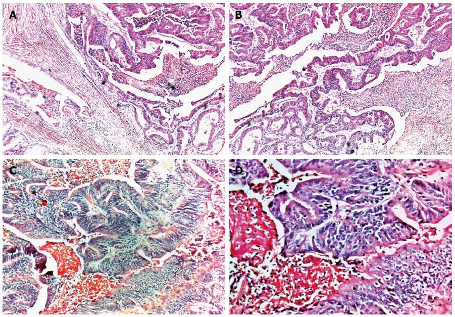
Figure 3 Histological findings of surgical specimen demonstrating moderately differentiated adenocarcinoma, infiltrating the wall thickness (A) (HE, magnification × 5), with areas of cribriform appearance due to fusion of glands and areas of necrosis (B) (HE, magnification × 10); A higher magnification, demonstrating dysplastic aspect of epithelium, loss of polarity and cell dysplasia (C) (HE, magnification × 100) and (D) (HE, magnification × 200).
HE: Hematoxylin and eosin.
- Citation: Paquissi FC, Lima AHFBP, Lopes MFDNV, Diaz FV. Adenocarcinoma of the third and fourth portions of the duodenum: The capsule endoscopy value. World J Gastroenterol 2015; 21(31): 9437-9441
- URL: https://www.wjgnet.com/1007-9327/full/v21/i31/9437.htm
- DOI: https://dx.doi.org/10.3748/wjg.v21.i31.9437


