©The Author(s) 2015.
World J Gastroenterol. Aug 14, 2015; 21(30): 9175-9181
Published online Aug 14, 2015. doi: 10.3748/wjg.v21.i30.9175
Published online Aug 14, 2015. doi: 10.3748/wjg.v21.i30.9175
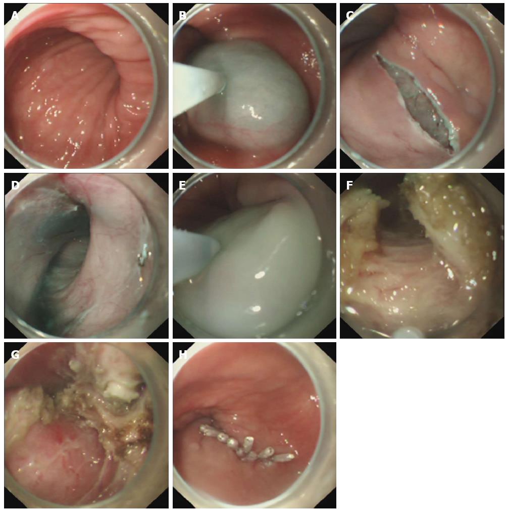
Figure 1 Case illustration of endoscopic full-thickness myotomy.
A: A twisting esophagus was shown before POEM; B: Submucosal injection was made to provide a cushion; C: A longitudinal mucosal incision was made as the tunnel entry; D: Submucosal tunnel creation; E: Submucosal injection to the esophageal cavity to preset the tunnel route; F-G: Endoscopic full-thickness myotomy, peri-esophageal membrane could be seen; H: The mucosal entry was closed with several clips. POEM: Peroral endoscopic myotomy.
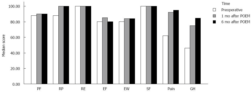
Figure 2 Quality of life outcomes (SF-36 domain).
- Citation: Li CJ, Tan YY, Wang XH, Liu DL. Peroral endoscopic myotomy for achalasia in patients aged ≥ 65 years. World J Gastroenterol 2015; 21(30): 9175-9181
- URL: https://www.wjgnet.com/1007-9327/full/v21/i30/9175.htm
- DOI: https://dx.doi.org/10.3748/wjg.v21.i30.9175


