©The Author(s) 2015.
World J Gastroenterol. Aug 14, 2015; 21(30): 9142-9149
Published online Aug 14, 2015. doi: 10.3748/wjg.v21.i30.9142
Published online Aug 14, 2015. doi: 10.3748/wjg.v21.i30.9142
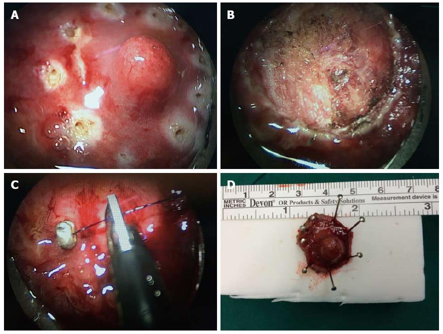
Figure 1 Full-thickness excision of a rectal neuroendocrine tumor visible under transanal endoscopic microsurgery.
A: The tumor and a clearance margin marked by coagulation dots using a needle electrode; B: After full-thickness excision with neat surgical margins; C: After suturing the rectal wall. The leakage from the suture line will be negligible; D: The neuroendocrine tumor removed completely with safe surgical margins.
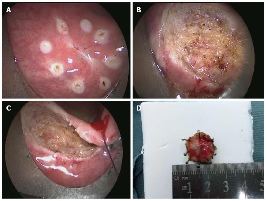
Figure 2 Full-thickness excision of a rectal lesion after incomplete resection.
A: The scar site after endoscopic polypectomy where the resection margin was identified as tumor positive; B: Fat tissue outside the rectal wall; C: Suturing of the rectal wall; D: Specimen obtained by transanal endoscopic microsurgery with a safe margin.
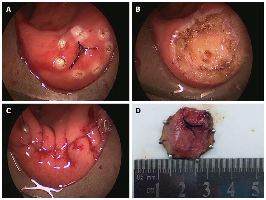
Figure 3 Full-thickness excision of an unobtrusive rectal neuroendocrine tumor using transanal endoscopic microsurgery.
A: The unobtrusive tumor found by trans-rectal ultrasound and marked by thread; B: The defect after full-thickness excision; C: Completion of suturing of the rectal wall; D: The neuroendocrine tumor removed completely with safe surgical margins.
- Citation: Chen WJ, Wu N, Zhou JL, Lin GL, Qiu HZ. Full-thickness excision using transanal endoscopic microsurgery for treatment of rectal neuroendocrine tumors. World J Gastroenterol 2015; 21(30): 9142-9149
- URL: https://www.wjgnet.com/1007-9327/full/v21/i30/9142.htm
- DOI: https://dx.doi.org/10.3748/wjg.v21.i30.9142


