©The Author(s) 2015.
World J Gastroenterol. Jan 21, 2015; 21(3): 1001-1008
Published online Jan 21, 2015. doi: 10.3748/wjg.v21.i3.1001
Published online Jan 21, 2015. doi: 10.3748/wjg.v21.i3.1001
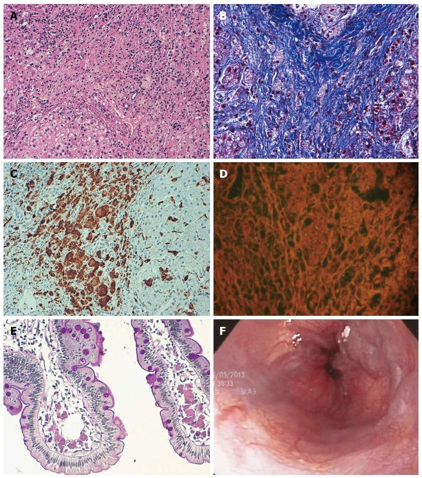
Figure 1 Liver histopathology and endoscopy images from sibling 1.
A: Presence of ballooning and pseudoacinar regeneration, with foamy cells (HE, magnification × 400); B: Presence of portal triads expanded by fibrosis tissue and foamy cells (Masson’s tricromic, magnification × 400); C: Positive CD68 immunoreactivity in cells (CD68 immunostaining); D: Autofluorescence of foamy cells in portal spaces (Masson’s tricromic, magnification × 400); E: Duodenal biopsy shows histiocytes with ceroid material in the lamina propria of the villi; F: Endoscopy showing esophageal varices.
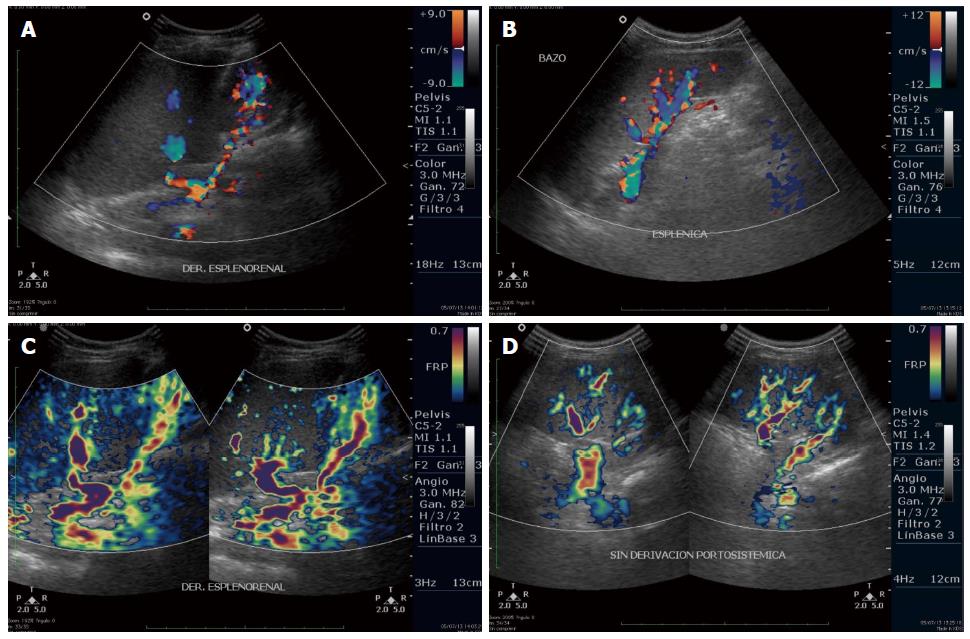
Figure 2 Comparison of Doppler ultrasound images from sibling 1 (A,C) and sibling 2 (B,D).
A: Splenic vein with a spontaneous splenorenal shunt; B: Splenic vein with collateral vessels at the hilum, without porto-systemic shunts; C: Dilated and tortuous vessels, collateral circulation and a spontaneous splenorenal shunt; D: Absence of a splenorenal shunt.
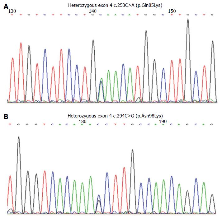
Figure 3 Electropherograms showing the mutations in exon 4 of the LIPA gene.
A: Mutation in exon 4: c.253C>A (p.Gln85Lys); B: Mutation in exon 4: c.294C>G (p.Asn98Lys).
-
Citation: Santillán-Hernández Y, Almanza-Miranda E, Xin WW, Goss K, Vera-Loaiza A, Mora MTGDL, Piña-Aguilar RE. Novel
LIPA mutations in Mexican siblings with lysosomal acid lipase deficiency. World J Gastroenterol 2015; 21(3): 1001-1008 - URL: https://www.wjgnet.com/1007-9327/full/v21/i3/1001.htm
- DOI: https://dx.doi.org/10.3748/wjg.v21.i3.1001


