Copyright
©The Author(s) 2015.
World J Gastroenterol. Jul 28, 2015; 21(28): 8580-8587
Published online Jul 28, 2015. doi: 10.3748/wjg.v21.i28.8580
Published online Jul 28, 2015. doi: 10.3748/wjg.v21.i28.8580
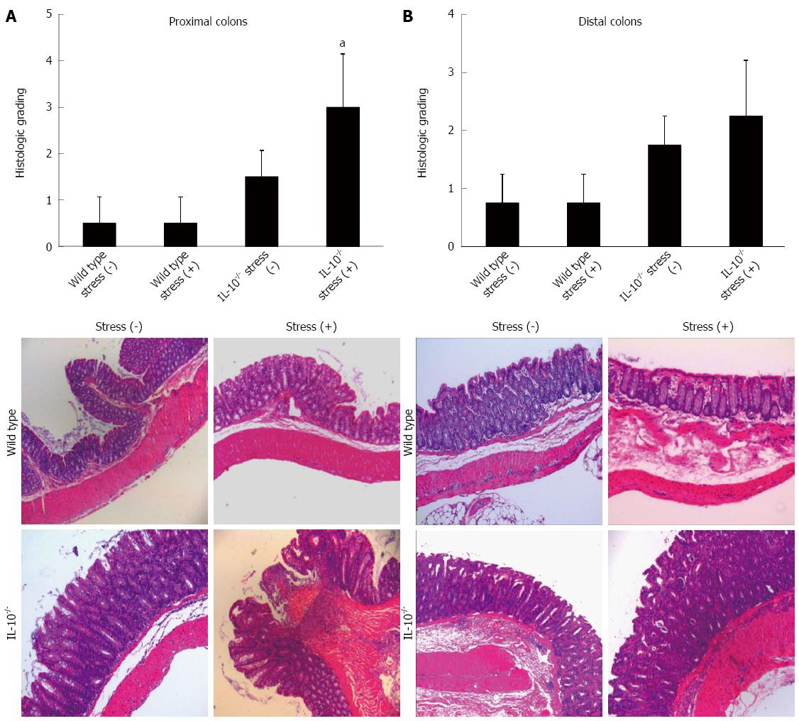
Figure 1 Histologic evaluations of (A) proximal and (B) distal colons in both wild-type and interleukin-10-/- mice.
Wild-type (stress positive, n = 4; stress negative, n = 4) and interleukin (IL)-10-/- mice (stress positive, n = 4; stress negative, n = 10) were physically restrained in a well-ventilated, 50cc conical polypropylene tube for 2 h per day for 3 consecutive days, as described in Materials and Methods. The total histologic score was derived from the severity of total inflammation and crypt damage (mean ± SD). Mild colitis was observed in some of IL-10-/- mice without restraint stress. In contrast, epithelial hyperplasia, crypt abscess, ulceration, and transmural inflammation were observed in IL-10-/- mice exposed to restraint stress. Results are representative of at least three separately examined sites (Magnification, x 40). Stress (+), restraint stress for 2 h; IL-10-/-: IL-10 knockout mice; aP < 0.05 vs stress negative IL-10-/- mice.
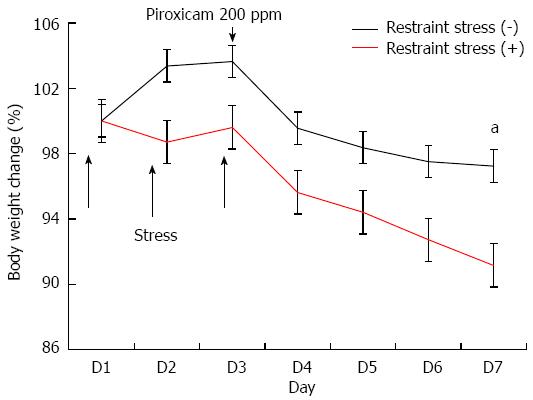
Figure 2 Body weight change in colitis model induced by piroxicam treatment.
Interleukin (IL)-10-/- mice (stress positive, n = 4; stress negative, n = 6) were exposed to restraint stress for 2 h per day for 3 consecutive days. IL-10-/- mice were then treated with piroxicam for 4 d at a dose of 200 ppm in the rodent chow. Body weight was evaluated daily and mice were sacrificed on the 7th day after the first exposure to restraint stress. Body weight was significantly reduced in IL-10-/- mice exposed to restraint stress 5 d after administration of piroxicam. aP < 0.05 vs stress negative IL-10-/- mice.
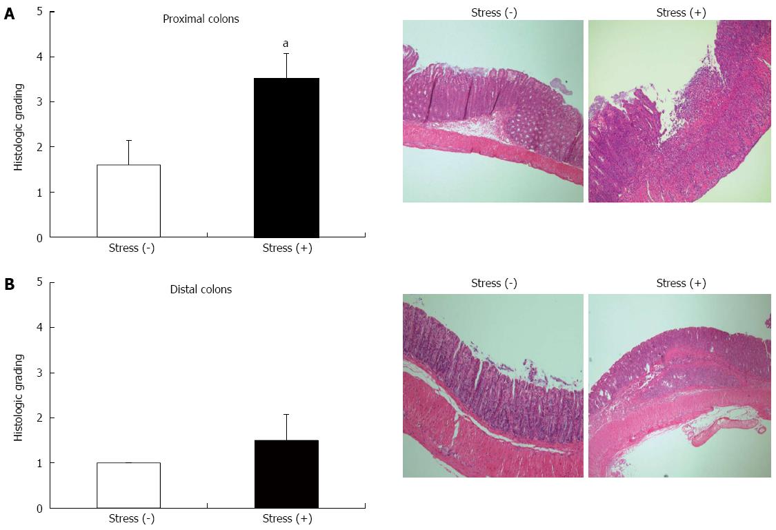
Figure 3 Histologic evaluations of (A) proximal and (B) distal colons in interleukin-10-/- mouse colitis induced by piroxicam.
Interleukin (IL)-10-/- mice were exposed to restraint stress for 2 h per day for 3 consecutive days. IL-10-/- mice were then treated with piroxicam for 4 d at a dose of 200 ppm in the rodent chow. The total histologic score was derived from the severity of total inflammation and crypt damage (mean ± SD). Mice with restraint stress exhibited severe colitis including inflammatory cell infiltration, crypt abscess, transmural inflammation, and ulceration, whereas mice without restraint stress showed epithelial hyperplasia and inflammatory cell infiltration. Results are representative of at least three separately examined sites (Magnification, x 40). Stress (+), restraint stress for 2 h. aP < 0.05 vs stress negative IL-10-/- mice.
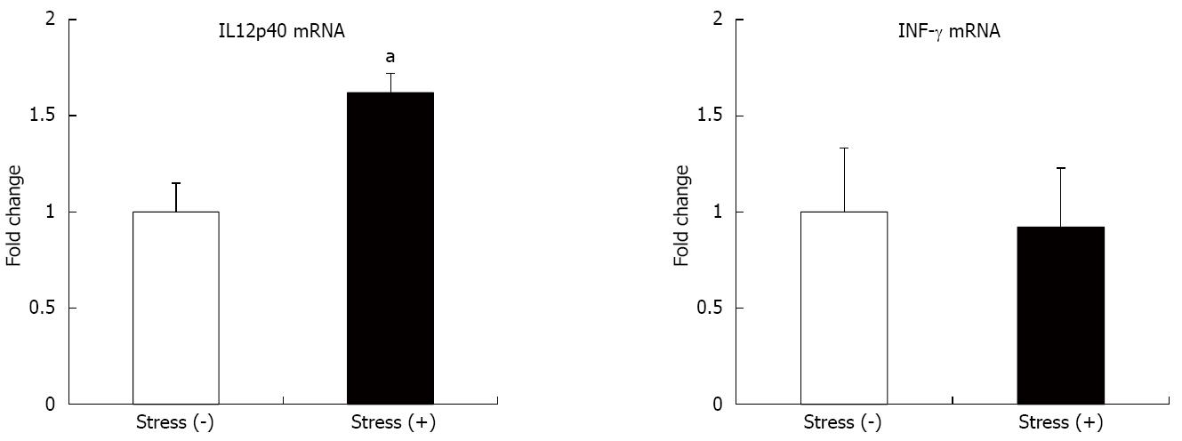
Figure 4 Restraint stress induced pro-inflammatory cytokine production in interleukin-10-/- mice with colitis.
Cytokine mRNA was extracted from mouse colon. Relative mRNA expression of interleukin (IL)-12p40 and IFN-γ against β-actin is expressed as the mean ± SE. The data are representative of three independent experiments. Stress (+), restraint stress for 2 h; aP < 0.05 vs stress negative IL-10-/- mice.
- Citation: Koh SJ, Kim JW, Kim BG, Lee KL, Kim JS. Restraint stress induces and exacerbates intestinal inflammation in interleukin-10 deficient mice. World J Gastroenterol 2015; 21(28): 8580-8587
- URL: https://www.wjgnet.com/1007-9327/full/v21/i28/8580.htm
- DOI: https://dx.doi.org/10.3748/wjg.v21.i28.8580


