©The Author(s) 2015.
World J Gastroenterol. Jul 14, 2015; 21(26): 8163-8169
Published online Jul 14, 2015. doi: 10.3748/wjg.v21.i26.8163
Published online Jul 14, 2015. doi: 10.3748/wjg.v21.i26.8163
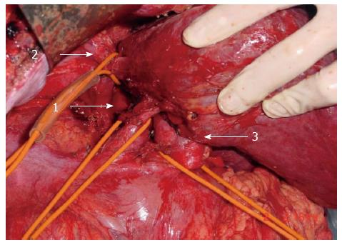
Figure 1 Inflow and outflow control during combined bilateral approach.
Control of infrahepatic inferior vena cava (IVC) (arrow 1), suprahepatic IVC (arrow 2), and hepatoduodenal ligament (arrow 3) during bilateral approach for ruptured hepatocellular carcinoma repair.
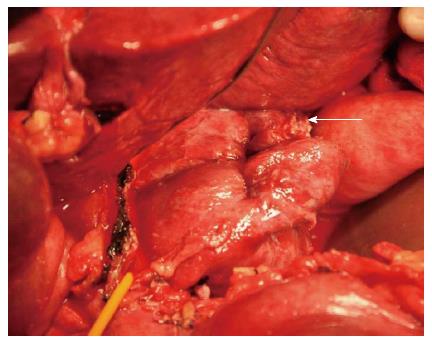
Figure 2 Rupture site access during left-sided approach.
Left-sided approach for ruptured hepatocellular carcinoma repair in Spiegel lobe. Rupture site is shown (white arrow).
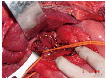
Figure 3 Anatomy of caudate lobe fossa and inferior vena cava.
Caudate lobe fossa (circumscribed by yellow dotted zone) and inferior vena cava (arrow) following removal of caudate lobe.
- Citation: Hong DF, Liu YB, Peng SY, Pang JZ, Wang ZF, Cheng J, Shen GL, Zhang YB. Management of hepatocellular carcinoma rupture in the caudate lobe. World J Gastroenterol 2015; 21(26): 8163-8169
- URL: https://www.wjgnet.com/1007-9327/full/v21/i26/8163.htm
- DOI: https://dx.doi.org/10.3748/wjg.v21.i26.8163


