©The Author(s) 2015.
World J Gastroenterol. Jul 14, 2015; 21(26): 8148-8155
Published online Jul 14, 2015. doi: 10.3748/wjg.v21.i26.8148
Published online Jul 14, 2015. doi: 10.3748/wjg.v21.i26.8148
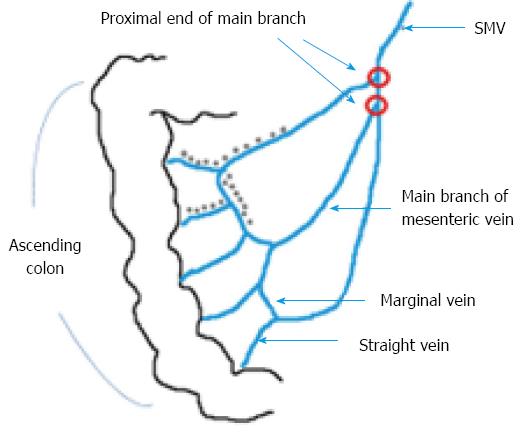
Figure 1 Schematic figure for calculating calcification score.
Calcifications (grey dots) at two straight veins, one marginal vein, and one main branch of mesenteric vein are noted. The proximal ends of main branches of mesenteric veins are not involved. Therefore, the calcification score in this case is calculated as 2 × 1 + 1 × 2 + 1 × 3 + 0 × 4 = 7.
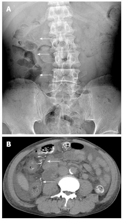
Figure 2 Kidney-ureter-bladder radiography and non-contrast enhanced computed tomography.
A 56-year-old male suffered from right upper quadrant abdominal pain and fever. A: The kidney-ureter-bladder radiography showed threadlike calcifications (arrows) at right abdomen; B: The non-contrast enhanced computed tomography study revealed calcifications (arrows) at tributaries of mesenteric vein and wall thickening of the ascending colon. The diagnosis is phlebosclerotic colitis with active episode.
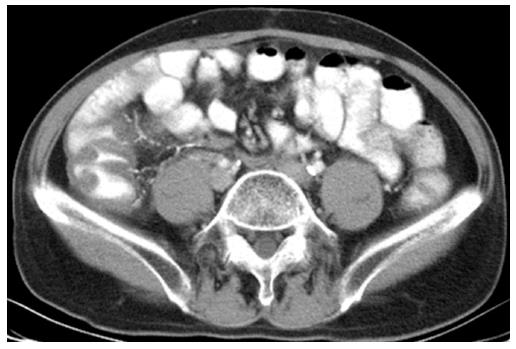
Figure 3 Intravenous contrast enhanced computed tomography with oral contrast ingestion.
A 59-year-old male came to emergency room with fever and tenderness at right lower abdomen. The contrast enhanced computed tomography shows wall thickening at the ascending colon and calcifications at straight veins, marginal veins and main branch of mesenteric vein, suggestive of phlebosclerotic colitis.
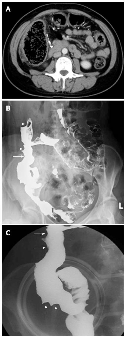
Figure 4 Barium follow-through study and intravenous contrast enhanced computed tomography.
A-54-year old female suffered from abdominal pain and constipation. A: The contrast enhanced abdominal computed tomography shows calcifications at tributaries of mesenteric vein and dilatation of the ascending colon; B: The barium follow-through study done after discharge shows thumb-printing appearance (arrows) at the ascending colon; C: The close view of cone compression.
Figure 5 Colonoscopy and plain-film radiographs of the abdomen of one symptomatic patient.
A: Cobble stone appearance in colonoscopy was noted; B: A polyp in the ascending colon was noted and biopsy was done. The pathology report revealed granulation tissue with heavy chronic inflammatory cell infiltration; C: The kidney-ureter-bladder radiography shows threadlike calcifications (arrows) at right abdomen with bowel obstruction.
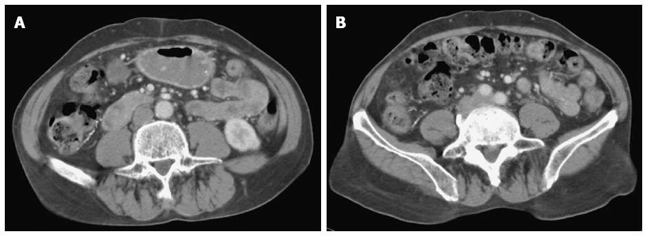
Figure 6 Intravenous contrast enhanced computed tomography of an asymptomatic patient.
A 72-year-old male has not experienced any significant abdominal symptoms. A: Computed tomography study showed calcifications at mesenteric veins; B: There was thickening of the wall of the cecum and ascending colon with pericolic stranding, which may indicate active disease.
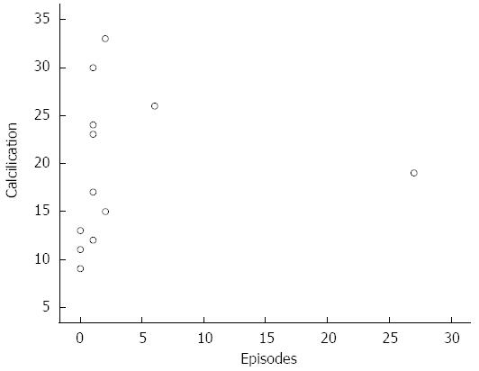
Figure 7 The dispersion diagram between the number of active disease episodes and the severity of mesenteric venous calcifications.
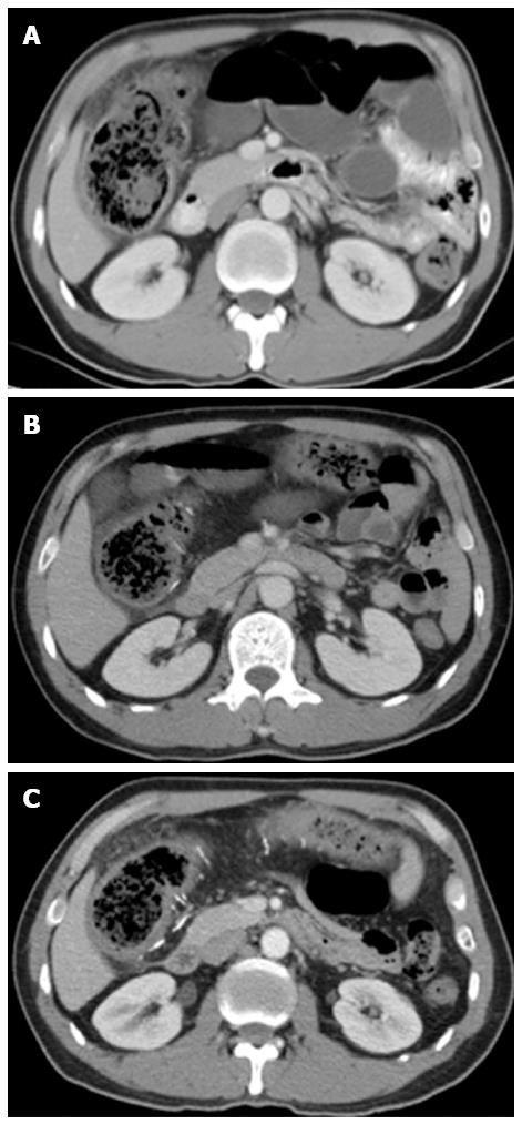
Figure 8 Serial computed tomography studies from 2005 to 2012.
A 46-year-old male suffered from recurrent abdominal pain and severe constipation for about 10 years. A: Computed tomography (CT) study done on November 18, 2005 revealed dilated ascending colon and minimal colonic wall thickening without evidence of mesenteric venous calcification; B: CT study of the same case done on November 17, 2009 showed some linear calcifications at colonic wall. Dilated ascending colon with wall thickening was also noted; C: CT study of the same case done on January 3, 2012 showed more linear calcifications at colonic wall and mesenteric veins. The extent of colonic wall thickening was also more prominent in this study.
- Citation: Yen TS, Liu CA, Chiu NC, Chiou YY, Chou YH, Chang CY. Relationship between severity of venous calcifications and symptoms of phlebosclerotic colitis. World J Gastroenterol 2015; 21(26): 8148-8155
- URL: https://www.wjgnet.com/1007-9327/full/v21/i26/8148.htm
- DOI: https://dx.doi.org/10.3748/wjg.v21.i26.8148


