©The Author(s) 2015.
World J Gastroenterol. Jul 14, 2015; 21(26): 8073-8080
Published online Jul 14, 2015. doi: 10.3748/wjg.v21.i26.8073
Published online Jul 14, 2015. doi: 10.3748/wjg.v21.i26.8073
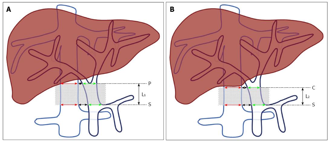
Figure 1 Effective length of the portacaval anastomosis using the magnetic compression technique.
A: When the origin of the portal vein (PV) branch is below the lower edge of the caudate lobe, the effective length, L1, is the distance between the intersection of the splenic vein and the PV, here S, and the origin of the PV branching, P. B: When the origin of the PV branch is higher than the lower edge of the caudate lobe, the effective length, L2, is the distance between S and the lower edge of caudate lobe, C. The red arrow indicates the diameter of the inferior vena cava (IVC); the green arrow indicates the diameter of the PV; the black arrow indicates the distance between the PV and the IVC.
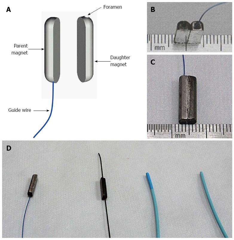
Figure 2 Magnetic compression system devices.
A: Schematic of the parent and daughter magnets; B, C: The magnetic compression system devices; D: Guide wire and catheter used to complete the operation.
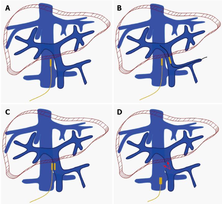
Figure 3 Surgical protocol and procedures.
A: The parent magnet was guided to the target position of the inferior vena cava (IVC) by the wire fixed to one end and the catheter; B: The daughter magnet was advanced to the anastomosis position of the portal vein (PV) by the guide wire and catheter through the incision on the splenic vein; C: Position of the magnets and the wire after the first surgery; D: The magnets were pulled out of the body by the wire fixed to the parent magnet and the portal-IVC shunt was set up.
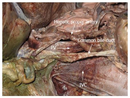
Figure 4 Anatomy of portacaval anastomosis in a cadaver.
The right posterior wall of the portal vein (PV) faces the left anterior wall of the inferior postcava. The common bile duct is at the right anterior of the PV. There is no important tissue in the postcaval space.
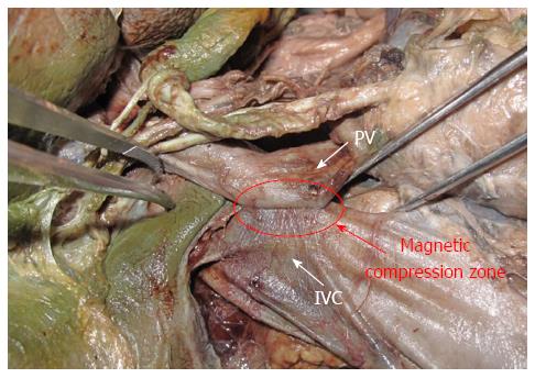
Figure 5 Compression of the walls of the portal vein and postcava implemented through the attraction of the parent and daughter magnets in a cadaver.
The elliptical area circled in red indicates the effective zone of the compression. No other vessel or bile duct was compressed. PV: Portal vein; IVC: Inferior vena cava.
-
Citation: Yan XP, Liu WY, Ma J, Li JP, Lv Y. Extrahepatic portacaval shunt
via a magnetic compression technique: A cadaveric feasibility study. World J Gastroenterol 2015; 21(26): 8073-8080 - URL: https://www.wjgnet.com/1007-9327/full/v21/i26/8073.htm
- DOI: https://dx.doi.org/10.3748/wjg.v21.i26.8073


