©The Author(s) 2015.
World J Gastroenterol. Jun 28, 2015; 21(24): 7594-7597
Published online Jun 28, 2015. doi: 10.3748/wjg.v21.i24.7594
Published online Jun 28, 2015. doi: 10.3748/wjg.v21.i24.7594
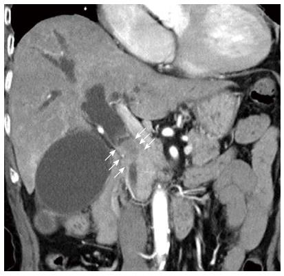
Figure 1 Contrast-enhanced computed tomography showed a 3-cm enhanced mass in the middle bile duct (white arrow) and dilatation of the intra-hepatic bile duct.
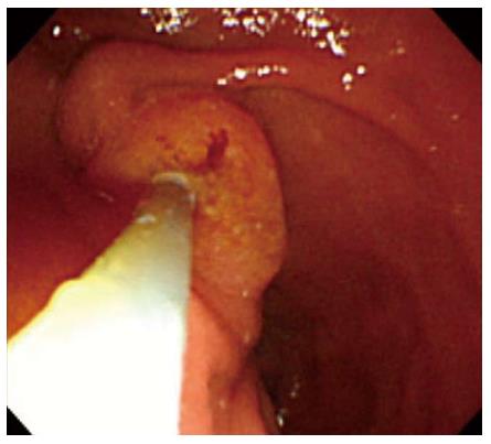
Figure 2 Endoscopic view of the papilla of Vater obtained by looking up from the anal side of the duodenal second portion.
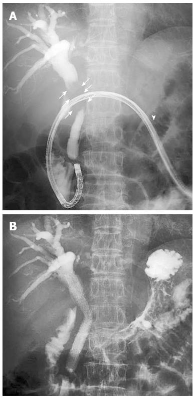
Figure 3 Cholangiogram showing a 3-cm stricture in the middle bile duct (white arrow); the scope was inserted through the gastric stoma (white arrowhead) (A), and successful placement of the 6F introducer metallic stent into the middle bile duct (B).
- Citation: Matsumoto K, Kato H, Tsutsumi K, Akimoto Y, Uchida D, Tomoda T, Yamamoto N, Noma Y, Horiguchi S, Okada H, Yamamoto K. Successful biliary drainage using a metal stent through the gastric stoma. World J Gastroenterol 2015; 21(24): 7594-7597
- URL: https://www.wjgnet.com/1007-9327/full/v21/i24/7594.htm
- DOI: https://dx.doi.org/10.3748/wjg.v21.i24.7594


