Copyright
©The Author(s) 2015.
World J Gastroenterol. Jan 14, 2015; 21(2): 556-562
Published online Jan 14, 2015. doi: 10.3748/wjg.v21.i2.556
Published online Jan 14, 2015. doi: 10.3748/wjg.v21.i2.556
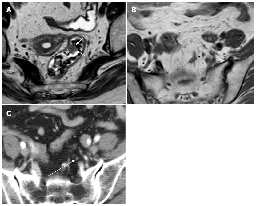
Figure 1 Early rectal carcinoma and lymph node metastasis on computed tomography and magnetic resonance imaging.
A: Axial T2-weighted image shows polypoid rectal carcinoma (arrows); B: Axial T1-weighted image shows regional lymph node with 4.5 mm in short axis diameter (black arrow); C: Axial computed tomography scan shows the same regional lymph node (white arrow) as in B.
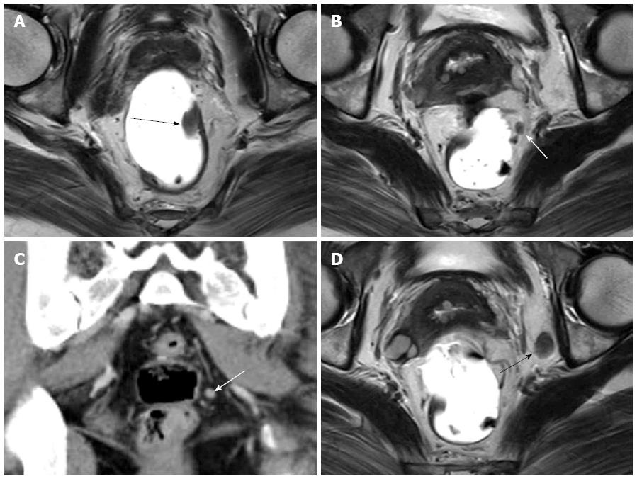
Figure 2 Early rectal carcinoma and lymph node metastasis on computed tomography and magnetic resonance imaging.
A: Axial T2-weighted image shows polypoid rectal carcinoma (black arrow); B: Axial T2-weighted image shows perirectal lymph node (white arrow); C: Coronal computed tomography scan shows the same lymph node (white arrow); D: Axial T2-weighted image shows an enlarged left obturator lymph node (black arrow). These metastatic lymph nodes were one-to-one correlated pathologically.
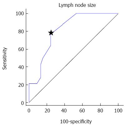
Figure 3 Receiver operating characteristic curve of short axis diameter.
A criterion of 4.1 mm (star) showed optimal sensitivity (78.6%) and specificity (75%).
- Citation: Choi J, Oh SN, Yeo DM, Kang WK, Jung CK, Kim SW, Park MY. Computed tomography and magnetic resonance imaging evaluation of lymph node metastasis in early colorectal cancer. World J Gastroenterol 2015; 21(2): 556-562
- URL: https://www.wjgnet.com/1007-9327/full/v21/i2/556.htm
- DOI: https://dx.doi.org/10.3748/wjg.v21.i2.556


