©The Author(s) 2015.
World J Gastroenterol. May 14, 2015; 21(18): 5744-5748
Published online May 14, 2015. doi: 10.3748/wjg.v21.i18.5744
Published online May 14, 2015. doi: 10.3748/wjg.v21.i18.5744
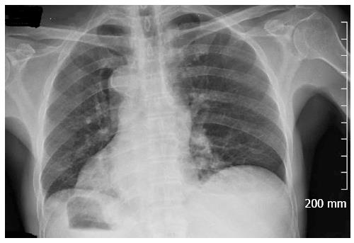
Figure 1 Chest X-ray revealed the tip of the heart was at the right side.
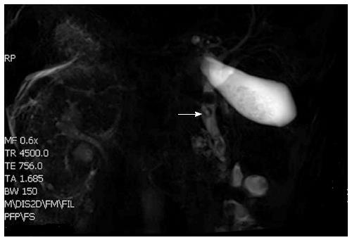
Figure 2 Total situs inversus viscerum in the abdomen and multiple stones in the gallbladder and common bile duct.
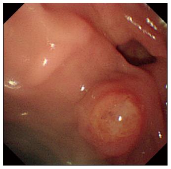
Figure 3 Duodenal diverticulum with an ampulla on its edge.
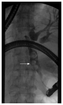
Figure 4 Cholangiography showed dilatation of the common bile duct and stones up to 12 mm with filling defects of the bile duct.
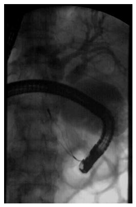
Figure 5 Multiple stones were removed with a Dormia basket.
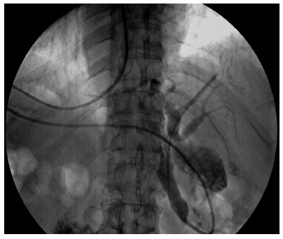
Figure 6 Cholangiography showed that there were no common bile duct stones.
- Citation: Hu Y, Zeng H, Pan XL, Lv NH, Liu ZJ, Hu Y. Therapeutic endoscopic retrograde cholangiopancreatography in a patient with situs inversus viscerum. World J Gastroenterol 2015; 21(18): 5744-5748
- URL: https://www.wjgnet.com/1007-9327/full/v21/i18/5744.htm
- DOI: https://dx.doi.org/10.3748/wjg.v21.i18.5744


