©The Author(s) 2015.
World J Gastroenterol. May 14, 2015; 21(18): 5513-5523
Published online May 14, 2015. doi: 10.3748/wjg.v21.i18.5513
Published online May 14, 2015. doi: 10.3748/wjg.v21.i18.5513
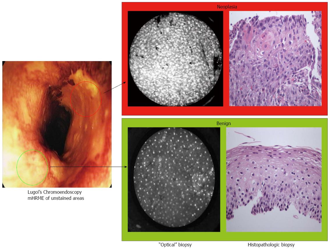
Figure 1 Lugol’s iodine unstained areas (high-resolution microendoscopy and optical biopsy vs tissue biopsy).
Lugol’s iodine unstained (abnormal) areas are imaged with high-resolution microendoscopy and optical biopsy and corresponding tissue biopsy of the area. Of the two unstained areas, only the sample presented in the upper panel was neoplastic (as characterized by loss of normal architecture and crowded nuclei).
Figure 2 Simplified model schematic of natural history.
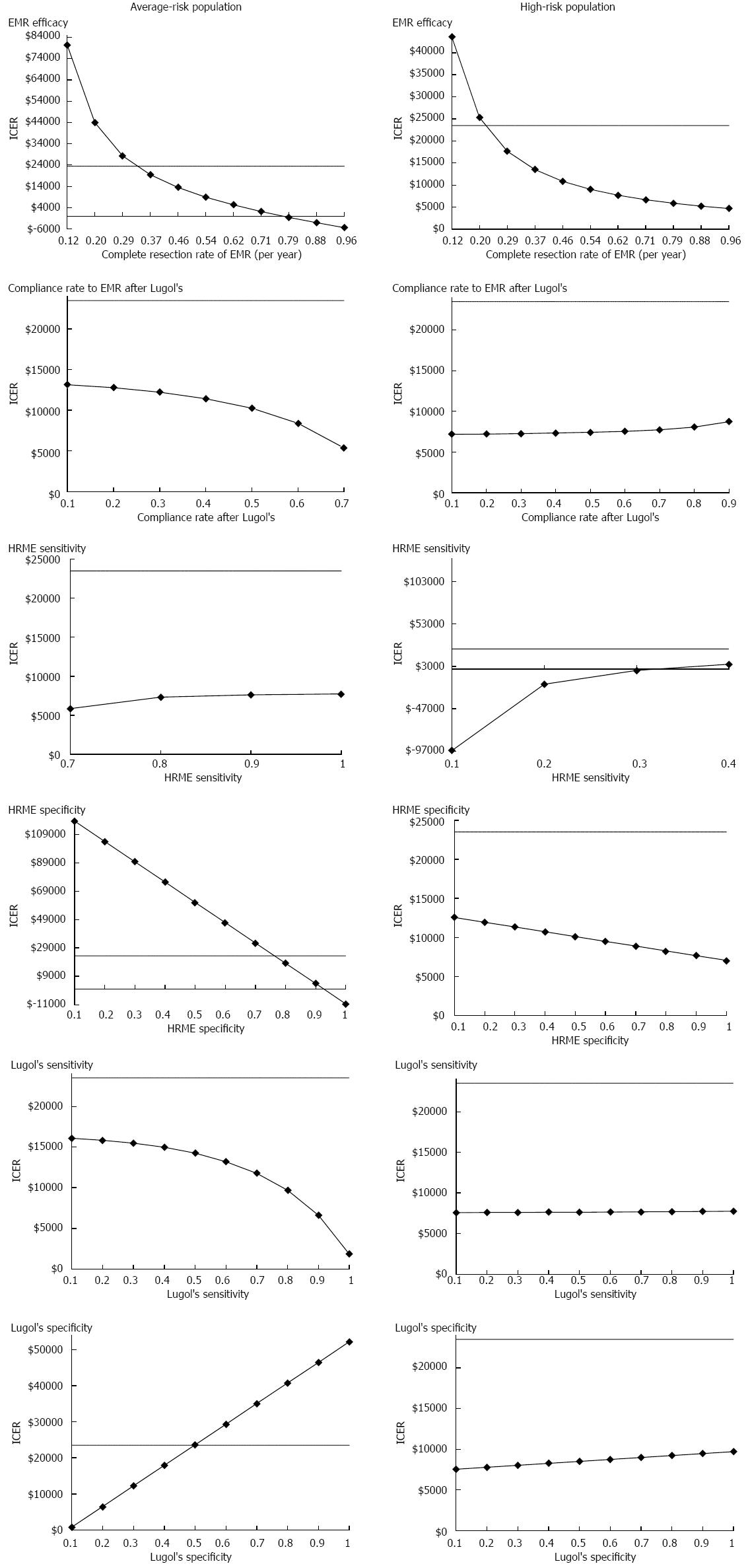
Figure 3 One-way sensitivity analyses.
EMR: Endoscopic mucosal resection; HRME: High-resolution microendoscopy; ICER: Incremental cost-effectiveness ratio.
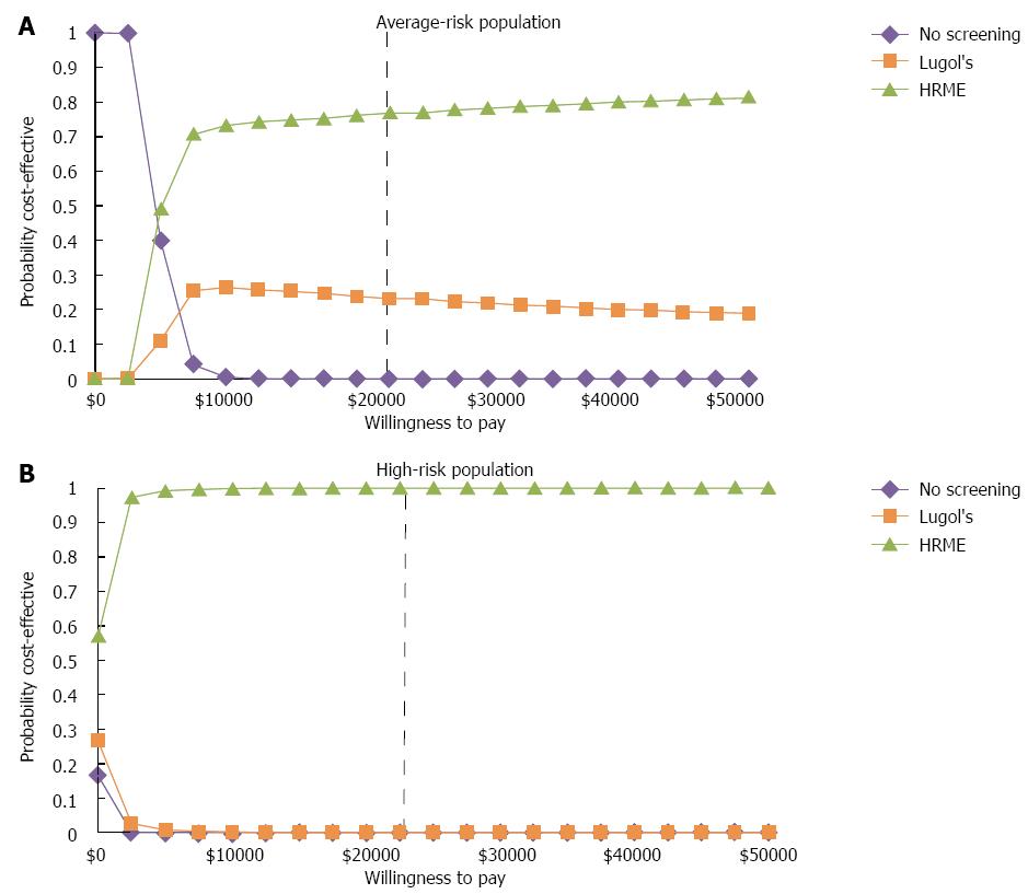
Figure 4 Probabilistic sensitivity analyses.
HRME: High-resolution microendoscopy.
- Citation: Hur C, Choi SE, Kong CY, Wang GQ, Xu H, Polydorides AD, Xue LY, Perzan KE, Tramontano AC, Richards-Kortum RR, Anandasabapathy S. High-resolution microendoscopy for esophageal cancer screening in China: A cost-effectiveness analysis. World J Gastroenterol 2015; 21(18): 5513-5523
- URL: https://www.wjgnet.com/1007-9327/full/v21/i18/5513.htm
- DOI: https://dx.doi.org/10.3748/wjg.v21.i18.5513


