Copyright
©The Author(s) 2015.
World J Gastroenterol. May 7, 2015; 21(17): 5311-5319
Published online May 7, 2015. doi: 10.3748/wjg.v21.i17.5311
Published online May 7, 2015. doi: 10.3748/wjg.v21.i17.5311
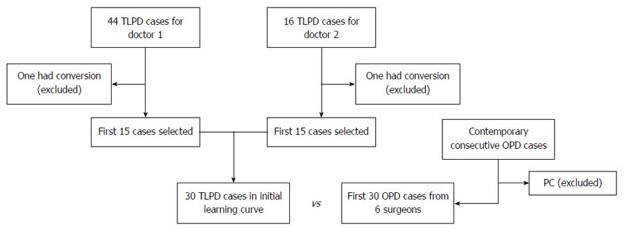
Figure 1 Flow chart of patient selection and exclusion.
TLPD: Total laparoscopic pancreaticoduodenectomy; OPD: Open pancreaticoduodenectomy; PC: Pancreatic cancer.
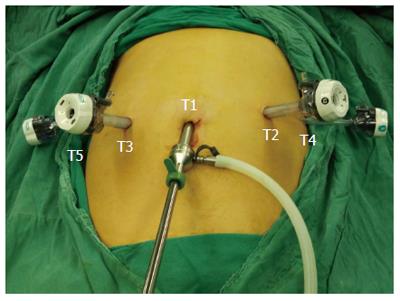
Figure 2 Placement of the 5 ports.
T1: 12-mm telescope trocar in the navel; T2: 12-mm trocar along the left midclavear line; T3: 12-mm trocar along the right midclavear line; T4: 5-mm trocar along the left anterior axillary line; T5: 5-mm trocar along the right anterior axillary line.
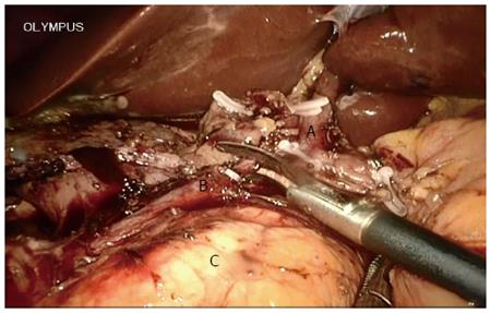
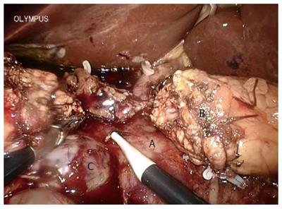
Figure 3 Gastroduodenal artery was ligated and divided.
A: Common hepatic artery; B: Gastroduodenal artery; C: Pancreas.
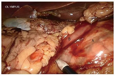
Figure 4 Mobilization of the duodenum using the Kocher maneuver.
A: Pancreatic head; B: Duodenum; C: Gallbladder.
Figure 5 Uncinate dissection.
A: Portal vein; B: Pancreatic stump; C: Cystic lesion in the uncinate.
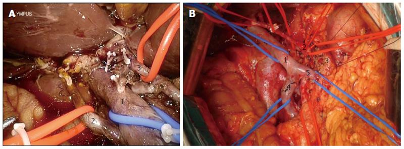
Figure 6 Skeletonization of the hepatoduodenal ligamentum.
A: During total laparoscopic pancreaticoduodenectomy (1: Portal vein; 2: Right hepatic artery; 3: Left hepatic artery); B: During open pancreaticoduodenectomy (1: Portal vein; 2: Common hepatic artery; 3: Pancreatic stump; 4: Superior mesenteric vein; 5: Splenic vein).
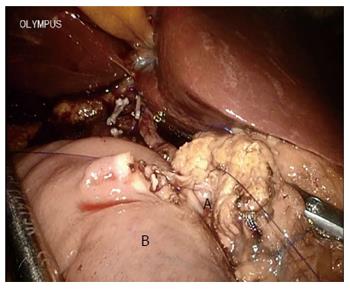
Figure 7 Modified mucosa-to-mucosa pancreaticojejunostomy.
A: Pancreatic duct; B: Jejunal stump.
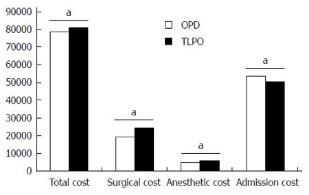
Figure 8 Cost analysis of the two cohorts.
aP < 0.05 vs TLPD. TLPD: Total laparoscopic pancreaticoduodenectomy; OPD: Open pancreaticoduodenectomy.
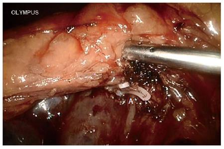
Figure 9 Small artery (A) below the pancreatic body caused bleeding when creating a tunnel between the superior mesenteric vein and the neck of the pancreas.
-
Citation: Tan CL, Zhang H, Peng B, Li KZ. Outcome and costs of laparoscopic pancreaticoduodenectomy during the initial learning curve
vs laparotomy. World J Gastroenterol 2015; 21(17): 5311-5319 - URL: https://www.wjgnet.com/1007-9327/full/v21/i17/5311.htm
- DOI: https://dx.doi.org/10.3748/wjg.v21.i17.5311


