Copyright
©The Author(s) 2015.
World J Gastroenterol. Apr 28, 2015; 21(16): 5110-5114
Published online Apr 28, 2015. doi: 10.3748/wjg.v21.i16.5110
Published online Apr 28, 2015. doi: 10.3748/wjg.v21.i16.5110
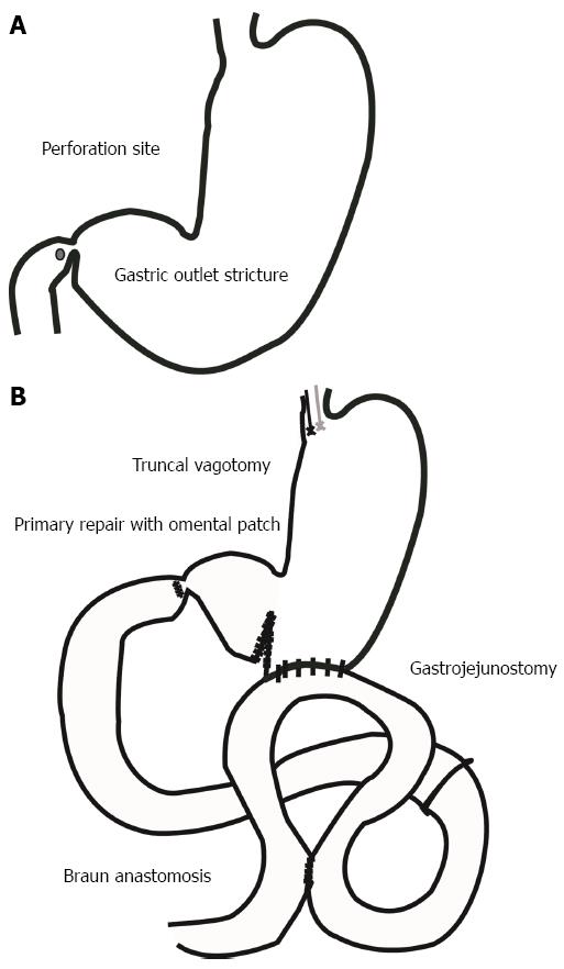
Figure 1 Schematic model of the operation.
A: Before the operation; B: The primary repair and omental patch, truncal vagotomy, gastrojejunostomy, and Braun anastomosis for the duodenal ulcer perforation and gastric outlet stricture.
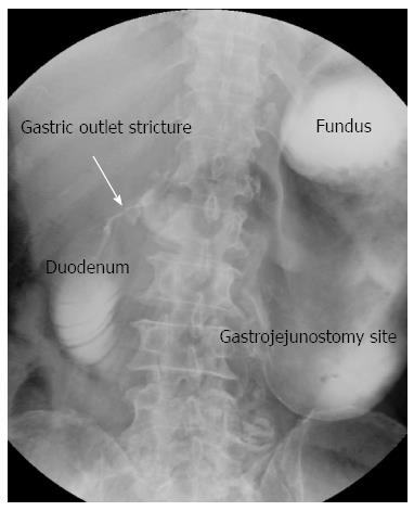
Figure 2 Upper gastrointestinal series finding on the 10th postoperative day.
Decreased peristalsis and severe distension of the stomach were observed as well as stricture on the duodenal perforation site. There was no visible contrast passage to the gastrojejunostomy.
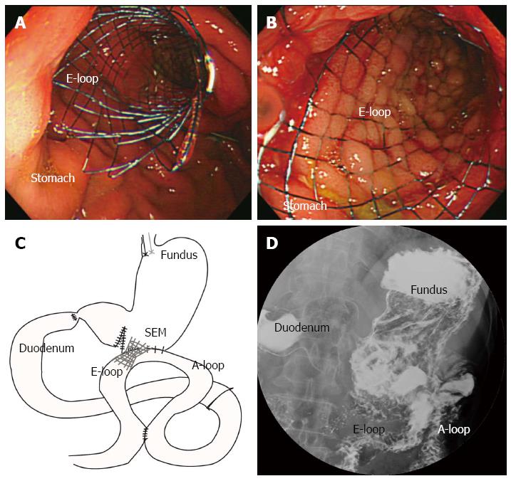
Figure 3 Endoscopic findings.
A, B: A Self-expandable metallic stent was inserted at the gastrojejunostomy anastomosis site over a guide wire; C: Schematic model of the post-insertion state; D: The upper gastrointestinal series finding after the stent insertion showing good passage of the contrast material from the esophagus to the jejunum, with no evidence of leakage at the anastomosis site. A-loop: Afferent loop; E-loop: Efferent loop.
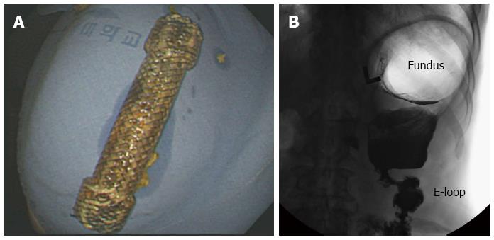
Figure 4 Follow-up upper gastrointestinal series.
A: Removal of the self-expandable metallic stent; B: The upper gastrointestinal series finding showed good passage of the contrast material to efferent loop (E-loop).
- Citation: Cha RR, Lee SS, Kim H, Kim HJ, Kim TH, Jung WT, Lee OJ, Bae KS, Jeong SH, Ha CY. Management of post-gastrectomy anastomosis site obstruction with a self-expandable metallic stent. World J Gastroenterol 2015; 21(16): 5110-5114
- URL: https://www.wjgnet.com/1007-9327/full/v21/i16/5110.htm
- DOI: https://dx.doi.org/10.3748/wjg.v21.i16.5110


