Copyright
©The Author(s) 2015.
World J Gastroenterol. Apr 28, 2015; 21(16): 5002-5008
Published online Apr 28, 2015. doi: 10.3748/wjg.v21.i16.5002
Published online Apr 28, 2015. doi: 10.3748/wjg.v21.i16.5002
Figure 1 Single charged-couple device integrated type endocytoscope (GIF-Y0002, a prototype from Olympus, Tokyo, Japan).
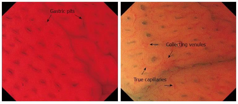
Figure 2 Normal pit-dominant type in the gastric corpus.
A regular arrangement of gastric pits is observed, with true capillary network.
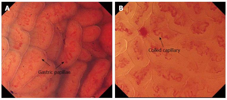
Figure 3 Normal papilla-dominant type in the gastric antrum (A) and the rounded papillae are tightly-packed and regularly arranged, surrounding coiled capillaries (B).
Figure 4 Irregular endocytoscopic patterns.
A: Unclear surface epithelium with necrotic tissue; B: Gastric pits (forming sulcus) are visualized when open and more easily stained; C: Completely stained epithelium, with goblet cells.
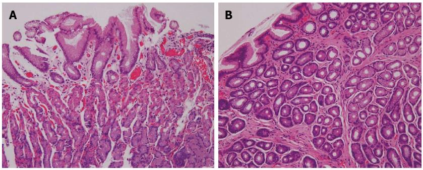
Figure 5 Histopathology of normal fundus gland and normal antral gland.
A: Histopathology showing the arrangement of normal fundus gland in the corpus. The arrangement of the surface mucosal epithelium is regular, with little inflammation; B: Histopathology showing the arrangement of normal antral gland. There is no inflammation and the arrangement of the surface mucosal epithelium is regular.
-
Citation: Sato H, Inoue H, Ikeda H, Sato C, Phlanusittepha C, Hayee B, Santi EGR, Kobayashi Y, Kudo SE.
In vivo gastric mucosal histopathology using endocytoscopy. World J Gastroenterol 2015; 21(16): 5002-5008 - URL: https://www.wjgnet.com/1007-9327/full/v21/i16/5002.htm
- DOI: https://dx.doi.org/10.3748/wjg.v21.i16.5002


