Copyright
©The Author(s) 2015.
World J Gastroenterol. Apr 21, 2015; 21(15): 4680-4687
Published online Apr 21, 2015. doi: 10.3748/wjg.v21.i15.4680
Published online Apr 21, 2015. doi: 10.3748/wjg.v21.i15.4680
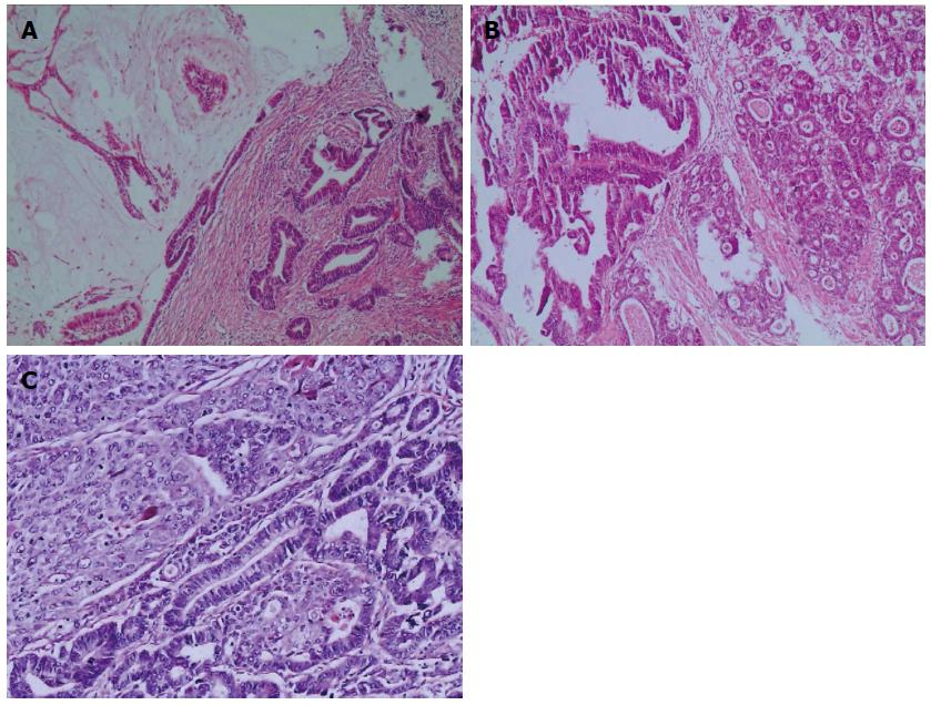
Figure 1 Hematoxylin and eosin staining of mixed gastric carcinoma.
A: Gastric mixed mucinous adenocarcinoma and tubular adenocarcinoma; B: Gastric mixed papillary adenocarcinoma and tubular adenocarcinoma; C: Gastric mixed tubular adenocarcinoma and squamous cell carcinoma (40 ×).
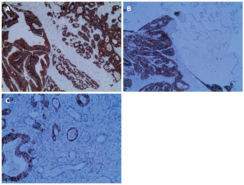
Figure 2 Human epidermal growth factor receptor 2 staining of mixed gastric carcinoma by immunohistochemistry.
A: Gastric mixed tubular adenocarcinoma and papillary adenocarcinoma showing extensive (HER2 3+) staining; B: Gastric mixed papillary adenocarcinoma and mucinous adenocarcinoma showing partial (HER2 2+); C: Gastric mixed carcinoma with tubular adenocarcinoma showing focal (HER2 1+) staining (40 ×). HER2: Human epidermal growth factor receptor 2.
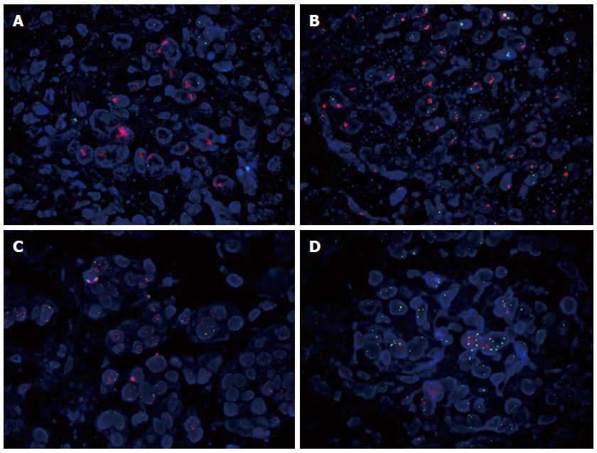
Figure 3 Human epidermal growth factor receptor 2 gene amplification and high copy number of chromosome 17 by fluorescence in situ hybridization method.
A: Gastric mixed carcinoma showing human epidermal growth factor receptor 2 (HER2) cluster amplification; B: Gastric mixed carcinoma showing HER2 large granule amplification; C: Gastric mixed carcinoma showing HER2 dot amplification. D. Gastric mixed carcinoma showing high polyploid 17; red signal = HER2 probe; green signal = chromosome 17 centromere probe (1000 ×). HER2: Human epidermal growth factor receptor 2.
- Citation: Wang YK, Chen Z, Yun T, Li CY, Jiang B, Lv XX, Chu GH, Wang SN, Yan H, Shi LF. Human epidermal growth factor receptor 2 expression in mixed gastric carcinoma. World J Gastroenterol 2015; 21(15): 4680-4687
- URL: https://www.wjgnet.com/1007-9327/full/v21/i15/4680.htm
- DOI: https://dx.doi.org/10.3748/wjg.v21.i15.4680


